Johny Marice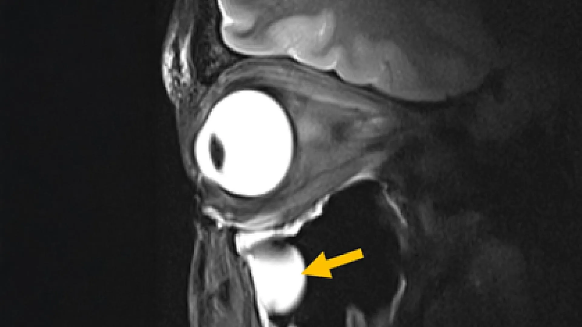
Magnetic resonance imaging (MRI) stands as a cornerstone of modern medical diagnostics, offering physicians an unparalleled non-invasive window into the human body. However, the inherent physics of MRI, particularly the transmission and reception of radiofrequency (RF) signals, presents significant challenges when attempting to generate high-resolution images of certain anatomical regions. Deep brain structures, the intricate tissues of the eye, and the surrounding orbital cavity have historically been difficult to visualize with the clarity and speed desired, often leading to prolonged examination times and potential diagnostic ambiguities. Now, a groundbreaking innovation from a collaborative team of researchers promises to revolutionize MRI technology, delivering sharper images in less time and expanding its diagnostic reach.
The pioneering work, led by Nandita Saha, a doctoral student within Professor Thoralf Niendorf’s Experimental Ultrahigh Field Magnetic Resonance laboratory at the Max Delbrück Center, has culminated in the development of a novel MRI antenna engineered with advanced metamaterials. This cutting-edge antenna not only produces significantly sharper images but also achieves this with reduced scanning durations. Crucially, its design is compatible with existing MRI systems, obviating the need for costly and disruptive upgrades to entire imaging machines. The findings detailing this significant advancement were recently published in the prestigious journal Advanced Materials.
This multidisciplinary endeavor brought together a diverse group of experts, pooling their knowledge from the fields of MRI physics, clinical ophthalmology, and translational imaging. The collaboration spanned institutions, including the Max Delbrück Center and Rostock University Medical Center, with researchers in Rostock actively contributing to the validation of this transformative technology for future clinical implementation.
Professor Thoralf Niendorf, the senior author of the research paper and a leading figure in ultrahigh-field MRI, articulated the fundamental principle behind their success. "By leveraging concepts derived from metamaterials, we have achieved a more efficient guidance of radiofrequency fields," Professor Niendorf explained. "This demonstrates a direct pathway where advanced physics can profoundly enhance medical imaging capabilities. This work charts a course toward faster, clearer MRI scans, offering substantial benefits to patients across a wide spectrum of clinical applications."
Metamaterials: A Paradigm Shift in MRI Antenna Design
The fundamental principle of MRI involves applying a powerful magnetic field and then transmitting RF signals into the body. Tissues within the body respond to these signals in specific ways, and the MRI scanner captures this information to construct detailed images. Generally, a stronger signal translates to clearer and more detailed diagnostic images. Traditional MRI antennas, often referred to as RF coils, have inherent limitations in their ability to effectively collect sufficient signal from tissues located deep within the body or from anatomically intricate regions. This often results in a compromise in image quality and necessitates longer scanning sessions to acquire adequate data.
The researchers’ breakthrough lies in their innovative integration of metamaterials directly into the MRI antenna. Metamaterials are not naturally occurring substances; instead, they are meticulously engineered structures designed to interact with electromagnetic waves in ways that defy the properties of conventional materials. In their experimental testing, the new metamaterial-enhanced antenna demonstrably amplified signals from targeted tissues, leading to a notable increase in spatial resolution, improved image sharpness, and accelerated data acquisition.
A significant advantage of this novel antenna is its seamless integration with current MRI equipment. This compatibility eliminates the substantial financial burden and logistical challenges associated with replacing entire MRI systems. The research team rigorously tested the antenna’s design by acquiring detailed images of the eye and orbit in human volunteers utilizing a 7.0 Tesla MRI scanner, a powerful system typically found in advanced research and clinical settings.
Professor Oliver Stachs, a co-author of the study and a prominent figure in ophthalmology at University Medicine Rostock, highlighted the immediate implications for his field. "Our research clearly demonstrates its relevance for ophthalmological applications," Professor Stachs stated. "It has the potential to facilitate anatomically detailed, high-spatial resolution MRI of the eye, offering an unprecedented view into the eye and its (patho)physiological processes that have historically remained largely inaccessible."
Beyond Ocular Imaging: Expanding the Horizon of MRI Applications
The ambition behind this project, as articulated by Nandita Saha, was to fundamentally re-evaluate MRI hardware through the lens of modern physics and antenna design principles. "Our goal was to rethink MRI hardware from the modern physics of antenna design," Saha elaborated.
Beyond its immediate impact on eye imaging, Saha indicated that the technology holds significant promise for other critical areas. The metamaterial antenna could be adapted to enhance patient safety during MRI examinations by mitigating unwanted heating effects around medical implants, a persistent concern for individuals with such devices. Furthermore, it could significantly improve the precision and efficacy of MRI-guided cancer treatments, such as tumor hyperthermia or thermal tissue ablation, by enabling more accurate targeting and delivery of RF energy.
Accelerated Diagnostics and Enhanced Patient Care
MRI examinations can be time-consuming and, at times, uncomfortable for patients, particularly when repeated scans are required due to difficulties in capturing crucial anatomical details. The new antenna’s ability to produce clearer images more rapidly has the potential to substantially shorten scan times. This not only improves patient comfort but also provides physicians with greater confidence in their diagnostic assessments, potentially leading to earlier and more accurate diagnoses.
The compact and lightweight nature of the antenna also offers an added benefit of enhanced patient comfort. Its customizable design allows it to be adapted for imaging various parts of the body, further improving the patient experience during MRI procedures.
Professor Niendorf expressed optimism about the technology’s future adaptability. He anticipates that the design can be further refined for MRI systems operating at magnetic field strengths both lower and higher than the 7.0 T scanner used in their initial tests. This broadens its applicability across a wider range of MRI scanners already in clinical use. Moreover, the antenna could be tailored for imaging organs beyond the eye, orbit, and brain, and could also play a crucial role in monitoring metabolic processes and tracking the pharmacokinetics of drug delivery within the body.
The potential also extends to specialized MRI techniques that focus on imaging atoms other than hydrogen, such as sodium and fluorine. By generating stronger signals and higher quality images for these less abundant nuclei, the metamaterial antenna could unlock new diagnostic insights in these advanced applications.
Dr. Ebba Beller, a co-author and researcher at Rostock University Medical Center, underscored the broader significance of this innovation. "Innovations in imaging hardware have the potential to transform diagnostics, and this study represents a crucial step towards the development of next-generation MRI technology," Dr. Beller commented.
Future Trajectories and Clinical Integration
The research team is actively preparing for the next phase of their work, which involves launching larger-scale clinical studies across multiple hospitals. Concurrently, they are modifying the antenna design to optimize it for imaging other vital organs, including the heart and kidneys. The established and productive collaboration between Professor Stachs and Professor Niendorf is set to continue, reinforced by reciprocal visiting scientist appointments, fostering ongoing knowledge exchange and joint research initiatives.
This significant project was made possible through funding from the German Research Foundation (DFG), operating as a joint initiative between the Max Delbrück Center and the Medical University Rostock. The successful integration of metamaterial science into MRI antenna design represents a pivotal moment in the evolution of medical imaging, promising a future of faster, clearer, and more comprehensive diagnostic capabilities for patients worldwide. The development signifies a tangible step forward from the fundamental physics laboratory to direct clinical impact, underscoring the power of interdisciplinary research in addressing pressing healthcare challenges. This advancement not only enhances current diagnostic accuracy but also lays the groundwork for entirely new avenues of medical investigation and treatment planning, potentially transforming patient outcomes across a multitude of diseases. The long-term implications suggest a future where MRI scans are not only more informative but also more accessible and less burdensome for the patient.