Researchers at the Massachusetts Institute of Technology (MIT) have made a groundbreaking discovery in optical physics, potentially revolutionizing the field of bioimaging. They have identified an unexpected phenomenon where a typically scattered and disordered laser signal, under precise conditions, can spontaneously reorganize into a highly focused, narrow "pencil beam." This novel capability promises to enable faster, more detailed, and less invasive imaging of living tissues, opening new avenues for understanding disease and evaluating therapeutic interventions.
The Genesis of a Scientific Surprise
The genesis of this remarkable finding can be traced back to experiments conducted by the team in the MIT Department of Electrical Engineering and Computer Science (EECS). Their prior work involved the development of a sophisticated "fiber shaper," a device meticulously engineered to exert precise control over laser light as it propagates through a multimode optical fiber. These fibers are chosen for their capacity to transmit substantial power levels, a critical factor in many advanced imaging techniques.
The research, detailed in a paper published today in the prestigious journal Nature Methods, began with a deviation from established theoretical expectations. "The common belief in the field is that if you crank up the power in this type of laser, the light will inevitably become chaotic," explained Sixian You, an assistant professor in EECS and a member of the Research Laboratory for Electronics, who served as the senior author on the paper. "But we proved that this is not the case. We followed the evidence, embraced the uncertainty, and found a way to let the light organize itself into a novel solution for bioimaging."
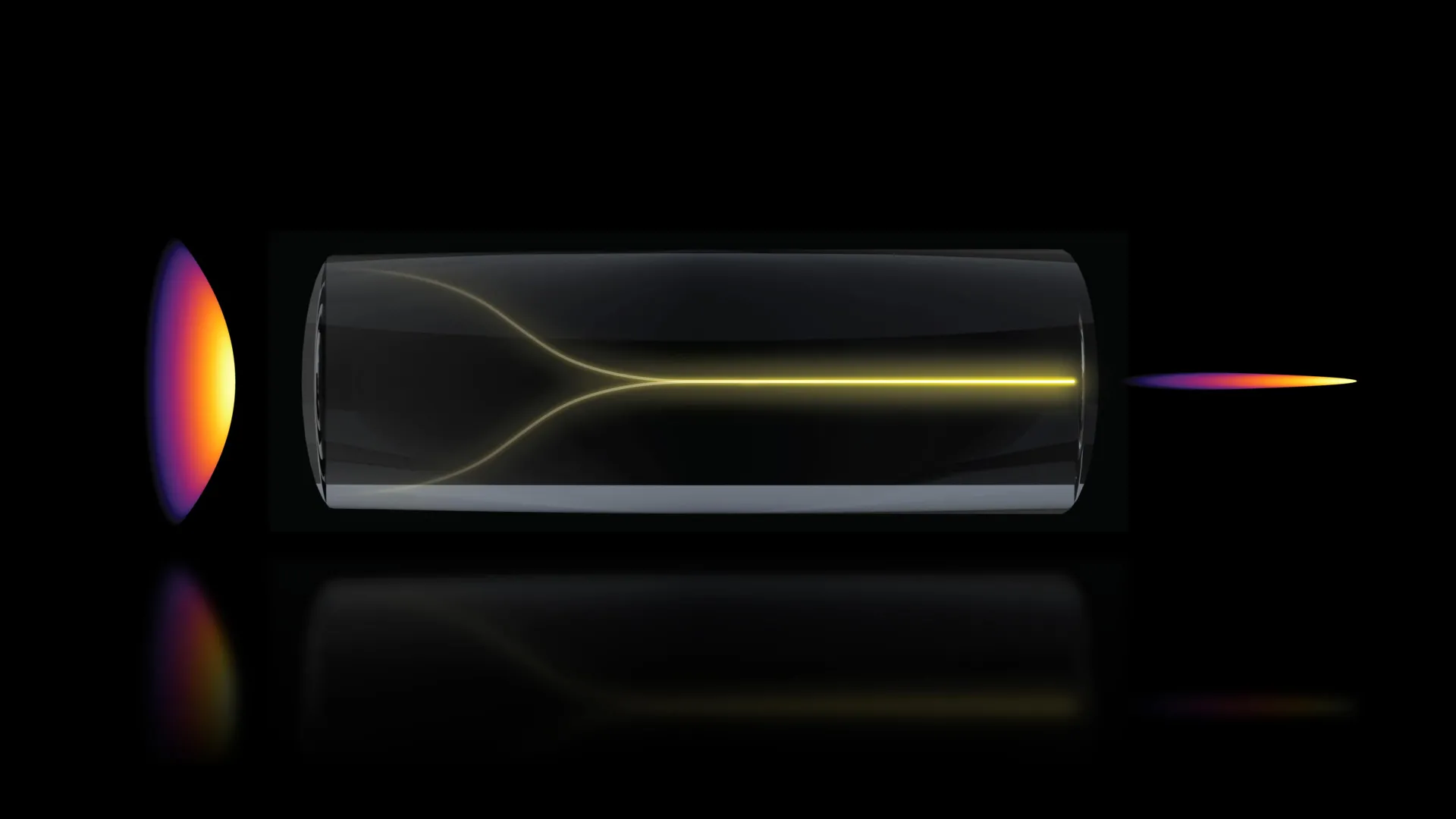
The lead author of the study, Honghao Cao, an EECS graduate student, was incrementally increasing the laser power to explore the operational limits of the fiber shaper. Standard optical theory predicts that as laser power intensifies within a multimode fiber, inherent imperfections in the fiber material lead to increased scattering and diffusion of the light. This typically results in a broad, less coherent beam. However, in this specific experimental setup, as the power approached a critical threshold – a level that would normally risk damaging the fiber – an astonishing transformation occurred. Instead of devolving into chaos, the laser light coalesced, sharpening into a single, extraordinarily focused beam resembling a pencil.
This unexpected self-organization bypasses the need for complex and often cumbersome external optical components that are traditionally employed to condition laser beams for imaging. "Disorder is intrinsic to these fibers," You elaborated. "The light engineering you typically need to do to overcome that disorder, especially at high power, is a longstanding hassle. But with this self-organization, you can get a stable, ultrafast pencil beam without the need for custom beam-shaping components."
Unlocking the Conditions for Self-Organization
The MIT team meticulously investigated the precise conditions that trigger this remarkable self-organizing behavior, identifying two pivotal requirements. The first and perhaps most stringent condition is the angle at which the laser beam enters the optical fiber. It must be aligned at a perfect, zero-degree angle relative to the fiber’s axis. This level of precision far exceeds the standard alignment protocols typically employed in optical experiments, which often allow for a degree of deviation.
The second crucial requirement involves power. The laser power must be elevated to a point where the light begins to interact directly with the glass material of the optical fiber itself. This interaction introduces a nonlinear effect, a phenomenon where the optical properties of the material change in response to the intensity of the light.
"At this critical power, the nonlinearity can counter the intrinsic disorder, creating a balance that transforms the input beam into a self-organized pencil beam," explained Cao. This delicate interplay between the inherent disorder of the multimode fiber and the emergent nonlinear optical properties of the glass at high power levels is the key to the phenomenon.
These specific conditions are rarely explored in conventional optical research. Scientists typically operate at lower power levels to safeguard delicate optical components, particularly the fibers, from damage. Furthermore, the inherent resilience of multimode fibers to accommodate a range of beam angles means that ultra-precise alignment is often deemed unnecessary. However, by deliberately combining these stringent requirements, the MIT researchers have unlocked a pathway to generate a stable, high-quality beam without resorting to elaborate optical engineering. "That is the charm of this method — you could do this with a normal, optical setup and without much domain expertise," You commented.
Enhanced Imaging Capabilities: Speed, Detail, and Reduced Artifacts
The self-formed pencil beam exhibits superior characteristics compared to beams generated through conventional methods. Rigorous testing revealed that this beam is exceptionally stable and possesses a remarkable level of detail. A common challenge in optical imaging is the presence of "sidelobes" – diffuse halos of light that emanate from the main beam, obscuring fine details and reducing overall image clarity. The self-organized pencil beam, however, remains remarkably clean and tightly focused, minimizing these artifacts and enabling sharper imaging.
To demonstrate the practical utility of this discovery, the researchers applied the technique to image the human blood-brain barrier (BBB). The BBB is a highly specialized and dense network of cells that forms a protective shield around the brain, preventing harmful substances from entering while simultaneously posing a significant obstacle for many therapeutic drugs. Understanding how drugs interact with and traverse this barrier is crucial for developing effective treatments for neurological disorders.
Revolutionizing the Visualization of the Blood-Brain Barrier
Traditional optical imaging methods used to study the BBB typically capture data in two-dimensional slices. To construct a comprehensive three-dimensional image, numerous individual scans are required, a process that is time-consuming and can introduce cumulative errors. This limitation has historically hampered the ability of researchers to observe dynamic processes, such as drug uptake by cells, in real time.
The new pencil beam approach, however, has dramatically accelerated this process. The MIT team was able to generate rapid, high-precision 3D images of the BBB, achieving imaging speeds approximately 25 times faster than current gold-standard techniques. Crucially, this speed increase did not come at the expense of image quality, which was preserved at a comparable level.
Beyond mere visualization, the method allows for the direct observation of individual cells absorbing drugs in real time. This capability is of immense interest to the pharmaceutical industry, which is actively seeking more accurate methods for screening drug candidates that can effectively cross the BBB. "The pharmaceutical industry is especially interested in using human-based models to screen for drugs that effectively cross the barrier, as animal models often fail to predict what happens in humans," stated Roger Kamm, the Cecil and Ida Green Distinguished Professor of Biological and Mechanical Engineering at MIT, and a co-author on the paper.
A particularly significant advantage of this new method is that it does not require the cells to be pre-tagged with fluorescent markers, a common prerequisite for many current imaging techniques. "For the first time, we can now visualize the time-dependent entry of drugs into the brain and even identify the rate at which specific cell types internalize the drug," Kamm added. This ability to observe the natural behavior of cells interacting with therapeutic compounds without artificial labeling provides a more accurate and physiologically relevant picture.
Sarah Spitz, a postdoc in the research group, further emphasized the broad applicability of the technique. "Importantly, however, this approach is not limited to the blood-brain barrier but enables time-resolved tracking of diverse compounds and molecular targets across engineered tissue models, providing a powerful tool for biological engineering," she remarked.
The researchers demonstrated that the system could produce cellular-level 3D images with enhanced quality and resolution. They also noted that the technique overcomes a common trade-off in microscopy, where improved resolution often comes at the cost of a reduced depth of focus. "Usually, you have a tradeoff between image resolution and depth of focus — you can only probe so far at a time," You explained. "But with our method, we can overcome this tradeoff by creating a pencil-beam with both high resolution and a large depth of focus."
Broader Implications and Future Directions
The implications of this discovery extend far beyond the immediate applications in blood-brain barrier research. The ability to rapidly and precisely image living tissues at a cellular level holds immense potential for advancing our understanding of a wide range of biological processes and diseases. For conditions such as Alzheimer’s disease and Amyotrophic Lateral Sclerosis (ALS), where the precise delivery and uptake of therapeutic agents in the brain are critical, this technology could offer unprecedented insights into treatment efficacy. Scientists could directly observe whether drugs are reaching their intended targets and how effectively cells are absorbing them, paving the way for more personalized and effective treatment strategies.
The research team is now focused on further elucidating the fundamental physics underpinning this self-organizing beam phenomenon. Understanding the precise mechanisms that govern its formation will be crucial for optimizing and expanding its applications. Future research plans include extending the method to image other complex biological structures, such as neural networks, and exploring pathways to translate this laboratory breakthrough into practical clinical and research tools.
The collaborative nature of this research is highlighted by the diverse group of scientists involved. Beyond You and Cao, the paper includes contributions from EECS graduate students Li-Yu Yu and Kunzan Liu, postdocs Sarah Spitz, Francesca Michela Pramotton, and Federico Presutti, Zhengyu Zhang (PhD ’24), Subhash Kulkarni (an assistant professor at Harvard University and the Beth Israel Deaconess Medical Center), and Roger Kamm.
This significant advancement was supported by a combination of funding sources, including MIT startup funds, the National Science Foundation (NSF), the Silicon Valley Community Foundation, the Diacomp Foundation, the Harvard Digestive Disease Core, a MathWorks Fellowship, and the Claude E. Shannon Award, underscoring the broad institutional and governmental recognition of its potential impact. As the research progresses, the self-organizing pencil beam promises to become an indispensable tool in the quest to unravel the complexities of life at the microscopic level and to develop the next generation of medical interventions.