Kang Muslim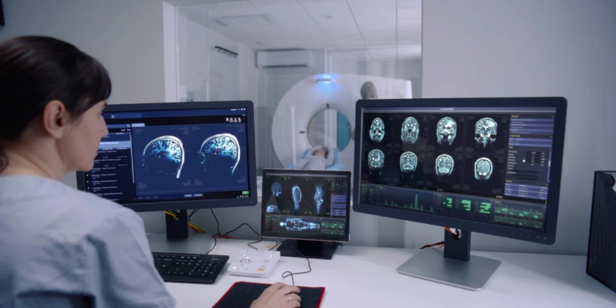
The quest to understand the biological foundations of human creativity has long occupied the intersection of psychology and neuroscience. While the ability to sketch or paint is a common human trait, the transition from hobbyist to professional artist involves a profound refinement of cognitive and motor skills. A recent study published in the journal Psychology of Aesthetics, Creativity, and the Arts has provided new insights into this transition, revealing that professional visual artists possess distinct patterns of brain structure and functional activity. These neural signatures appear to support the heightened mental imagery and complex motor coordination required to translate an internal vision into a tangible work of art.
Led by Erdem Taskiran of the University of Trento, the research marks a departure from traditional studies that focus on general creativity. By utilizing a "data fusion" approach—combining multiple types of brain imaging—the team has identified a comprehensive neural profile that distinguishes the professional artist from the layperson. This profile suggests that artistic expertise is not localized to a single "creative center" but is instead the result of a sophisticated integration of visual processing, motor control, and memory systems.
Methodology and the Multimodal Approach
The study’s design was rooted in the necessity of moving beyond single-measure neuroimaging. Historically, research into the "creative brain" has often relied on either structural MRI (looking at the size of brain regions) or functional MRI (looking at brain activity during tasks). However, Taskiran and his colleagues argued that artistic expertise is too complex to be captured by one dimension alone. To address this, they recruited 24 participants: 12 professional visual artists and 12 non-artists who served as a control group. The groups were carefully matched for age and gender to ensure that any observed differences were likely attributable to artistic training and practice rather than demographic variables.
The researchers employed three distinct types of magnetic resonance imaging (MRI). First, they used structural imaging to measure gray matter volume, which represents the density of neuronal cell bodies. Second, they utilized diffusion tensor imaging (DTI) to examine white matter pathways—the "wiring" of the brain that allows different regions to communicate. Finally, they recorded resting-state functional MRI to observe how different brain areas synchronize their activity when the participant is not performing a specific task.
By applying a machine learning algorithm to these datasets, the researchers were able to "fuse" the information. This allowed them to identify a single, multidimensional pattern that characterized the artist’s brain more accurately than any individual scan could.
The Structural Blueprint: Gray Matter and Visual Planning
One of the most significant findings of the study was the increased gray matter volume in professional artists within specific regions associated with high-level cognitive functions. These areas include parts of the prefrontal cortex, which is responsible for executive functions such as planning, decision-making, and complex problem-solving.
In the context of visual art, the prefrontal cortex is essential for the "pre-visualization" phase. An artist must not only imagine a scene but also plan the technical steps required to execute it—deciding on composition, perspective, and color theory before the brush even touches the canvas. The increased gray matter in these regions suggests a structural adaptation to the constant demand for strategic thinking in the creative process.
Furthermore, the study identified greater gray matter volume in regions associated with memory and visual processing. This aligns with the "expert" model of brain development seen in other fields, such as chess or music. Professional artists often possess a vast mental library of visual references and techniques. The structural enhancement of memory-related areas likely facilitates the rapid retrieval of these visual templates, allowing artists to manipulate and recombine them into original works.
The Neural Superhighway: Enhanced Connectivity
While gray matter represents the processing power of the brain, white matter represents the efficiency of communication between those processors. The study found that professional artists exhibited significantly stronger white matter connections, particularly in pathways linking the visual cortex to the executive control and motor centers of the brain.
This heightened connectivity is crucial for the "eye-to-hand" coordination that defines professional craftsmanship. In a non-artist, the path from perceiving a visual stimulus to executing a precise motor movement may be less direct or efficient. In a professional artist, the neural "highway" between the visual system (which processes the image) and the motor system (which controls the hand) is more robust. This allows for a more seamless translation of visual information into the fine motor movements required for drawing or painting.
The researchers also noted enhanced connectivity in pathways related to the "default mode network" (DMN). The DMN is typically active when the mind is wandering or engaged in internal thought. In artists, the integration of the DMN with executive networks may allow them to better harness spontaneous "flashes of inspiration" and channel them into structured, goal-oriented artistic production.
The Subcortical Engine: Cerebellum and Basal Ganglia
Perhaps the most novel aspect of the study was its focus on subcortical structures, specifically the cerebellum and the basal ganglia. For decades, these regions were primarily associated with basic motor control and habit formation. However, recent neuroscience has revealed their vital role in high-level cognition and learning.
The study found greater synchronization and activity within these regions in professional artists. The cerebellum is known to be involved in "internal models"—the brain’s ability to predict the outcome of a motor action before it happens. For an artist, this means the cerebellum helps "calculate" exactly how a stroke of charcoal will look based on the pressure and angle of the hand.
The basal ganglia, on the other hand, are central to the development of "procedural memory" or habits. Through years of practice, certain artistic techniques become second nature. The findings suggest that the artist’s brain has optimized these subcortical systems to handle the technical aspects of creation, thereby freeing up cortical resources for higher-level conceptual and aesthetic decisions. This shifts the understanding of creativity from a purely "intellectual" process to one that is deeply rooted in the brain’s motor and habit-learning circuitry.
Mental Imagery and the Mind’s Eye
A critical component of the research involved the Vividness of Visual Imagery Questionnaire (VVIQ). This psychometric tool requires participants to rate the clarity and detail of images they conjure in their minds. The results showed a direct correlation between the identified brain patterns and the reported vividness of mental imagery.
Professional artists consistently reported more "lifelike" and stable mental images compared to the control group. This suggests that the structural and functional differences observed in the MRI scans have a direct functional outcome: the ability to "see" a finished piece of art in the mind’s eye with high resolution. This internal visualization serves as a blueprint, guiding the artist through the long and often arduous process of creation.
Context and Chronology of Neuroaesthetics
The study by Taskiran and his colleagues arrives at a pivotal moment in the field of neuroaesthetics. The discipline, which began to gain traction in the late 1990s and early 2000s through the work of pioneers like Semir Zeki and V.S. Ramachandran, initially focused on how the brain perceives beauty. Early research often looked at the "reward centers" of the brain, such as the medial orbitofrontal cortex, which lights up when we see something we find aesthetically pleasing.
However, the focus has shifted over the last decade from perception to production. Researchers are no longer just asking "Why do we like art?" but "How do we make art?" This shift was marked by a series of studies on musicians and dancers, which established that long-term training leads to neuroplastic changes. The current study extends this timeline by providing one of the most detailed multimodal analyses of visual artists to date, moving the conversation beyond simple "right-brain" myths into a more nuanced understanding of whole-brain integration.
Implications for Education and Neuroplasticity
The findings of this study have significant implications for the fields of art education and cognitive rehabilitation. If artistic expertise is tied to specific neural patterns, it raises the question of how these patterns are formed. The concept of neuroplasticity suggests that the brain is not a static organ but is shaped by experience and practice.
For educators, this research reinforces the value of technical training. The involvement of the basal ganglia and cerebellum suggests that the "repetitive" aspects of art—such as practicing line work or color mixing—are not just about building manual dexterity; they are about restructuring the brain to automate technical tasks.
In a clinical context, the link between art and the brain’s motor and executive systems supports the use of art therapy for individuals with neurological conditions. By engaging the visual, motor, and planning centers of the brain simultaneously, artistic activity may help strengthen neural pathways that have been damaged by stroke or neurodegenerative diseases.
Limitations and the Nature vs. Nurture Debate
Despite the depth of the findings, the researchers have been careful to note the study’s limitations. The sample size of 24 individuals, while typical for intensive multimodal MRI studies, is relatively small. Larger-scale studies will be necessary to confirm if these patterns are universal across different types of visual artists, such as sculptors or digital animators.
Furthermore, the cross-sectional nature of the study—comparing artists and non-artists at a single point in time—leaves the question of causality open. It remains unclear whether the observed brain differences are the result of years of intense training (nurture) or whether individuals born with these specific neural configurations are more likely to be drawn to and succeed in professional art (nature).
The authors suggest that the reality is likely a combination of both. A biological predisposition toward vivid mental imagery might lead a child to spend more time drawing, and that continued practice then further shapes the brain’s structure and connectivity in a reinforcing loop.
Conclusion and Future Directions
The study by Taskiran et al. provides a compelling look into the "hardware" of the creative mind. By showing that professional expertise involves a widespread network including the cerebellum, sensorimotor systems, and subcortical structures, the research moves the scientific understanding of art away from the abstract and into the realm of concrete biological systems.
As neuroscience continues to advance, future research may look at how these neural patterns change over the course of an artist’s career, from the first years of training to the mastery of old age. For now, the "artist’s brain" stands as a testament to the human capacity for specialization, illustrating how the pursuit of beauty and expression can quite literally reshape the physical structure of the mind.