Kang Muslim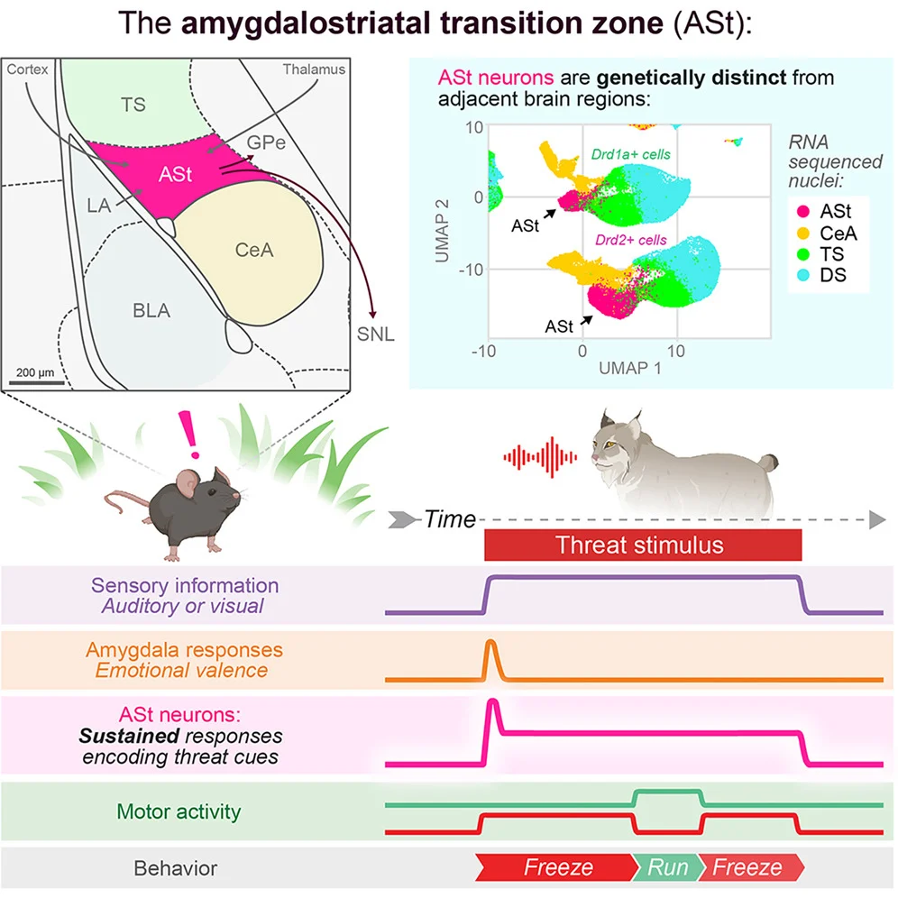
For decades, neuroscientists have faced a persistent paradox when studying how mammalian brains process fear and danger. While the amygdala—the brain’s recognized center for emotional processing—rapidly detects environmental threats, its electrical signals fade almost instantaneously, lasting merely milliseconds. Yet, when an animal or human encounters a hazard, physiological defensive behaviors such as freezing, hypervigilance, and evasive action persist for tens of seconds or even minutes. This temporal disconnect suggested that a crucial missing link existed within the neural circuitry, bridging the initial flash of threat recognition with sustained defensive action.
Groundbreaking research published in the journal Neuron has now identified that missing link. Led by Fergil Mills, an assistant professor at the University of Utah who conducted the work as a postdoctoral fellow in the laboratory of senior author Kay M. Tye at the Salk Institute for Biological Studies, a multi-institutional team of researchers has detailed the function of the amygdalostriatal transition zone (ASt). This largely unexplored cluster of brain cells acts as a continuous alarm signal generator, maintaining high-alert states during threats and redefining our fundamental understanding of neurobiology, fear conditioning, and neuropsychiatric disorders.
Anatomy of a Hidden Brain Region
Situated precisely between the amygdala and the striatum—a brain structure primarily responsible for controlling physical movements—the amygdalostriatal transition zone has historically escaped intensive scientific scrutiny. While neighboring regions received exhaustive mapping in relation to trauma, anxiety, and reward processing, the ASt remained an anatomical no-man’s-land.
To decipher the identity of this obscure region, the research team initiated a deep genetic profiling of the ASt and its surrounding tissues. By extracting and analyzing the genetic material of 97,434 individual cell nuclei from murine brain samples, the scientists uncovered a remarkably distinct genetic signature unique to the ASt.
The genomic analysis revealed that the ASt is heavily populated by a specific category of neurons known as Drd2-positive neurons, which express specialized receptors for dopamine, a key chemical messenger involved in motivation, learning, and response selection. Specifically, Drd2-positive neurons comprise approximately 71 percent of the total cellular makeup of the ASt. By contrast, a related cell type within the region, Drd1a-positive neurons, accounted for just 26 percent of the population. This overwhelming concentration of dopamine receptor-bearing cells provided the first major clue that the ASt possesses specialized properties suited for regulating complex behavioral outputs.
Historical Context and the Evolution of Fear Research
To fully appreciate the significance of the ASt discovery, neuroscientists must look back at decades of incremental breakthroughs in fear circuitry.
In 1995, a landmark study examining the brains of rats demonstrated that fear-learning neurons within the amygdala respond to threatening acoustic cues with rapid, fleeting bursts of electrical activity lasting only a fraction of a second. Years later, in 2002, subsequent laboratory investigations mapped the rapid transmission of these warning signals as they traveled directly from the amygdala into the neighboring, unmapped ASt.
However, a major theoretical puzzle emerged in 2009 through rodent research highlighting that amygdala signaling extinguished almost immediately upon threat presentation, despite the test subjects maintaining visible defensive behaviors—such as freezing—for extended periods. For nearly two decades, the neurological mechanism responsible for sustaining these prolonged survival responses remained unknown. The current study directly addresses this historical gap, confirming that the ASt transforms transient inputs from the amygdala into sustained, long-duration behavioral outputs.
Rigorous Testing and Real-Time Observation
To determine whether ASt neurons actively provide the continuous alarm signal required for extended defensive states, the researchers deployed an array of advanced neuroscientific techniques, including in vivo electrophysiology, optogenetics, and miniaturized head-mounted microscopy.
The team began by measuring the electrical activity of ASt neurons in 15 live mice undergoing behavioral conditioning. The animals were exposed to two distinct 20-second auditory cues: one that reliably predicted a mild foot shock, and another that signaled the delivery of a sweet liquid reward. Electrophysiological recordings revealed that ASt neurons responded selectively and robustly to the shock-predicting tone.
Unlike the transient, millisecond bursts characteristic of the amygdala, ASt neurons maintained continuous electrical firing for the entire 20-second duration of the threat cue. Furthermore, a direct correlation emerged between neural activity and behavior: during trials where mice exhibited the highest levels of freezing, ASt firing rates spiked to levels over three times higher than those observed during trials with minimal defensive movement.
To establish causality rather than mere correlation, the researchers employed optogenetics—a sophisticated method allowing scientists to control targeted populations of neurons using pulses of blue laser light. Testing 18 genetically engineered mice whose ASt neurons were photosensitive, the team observed that shining light directly into the ASt caused the animals to immediately freeze in place. When one side of an experimental enclosure was programmed to trigger the laser upon entry, the mice actively avoided that territory.
Further isolation experiments involving 35 additional mice pinpointed the exact cellular drivers of this behavior. Activating the Drd2-positive neurons replicated the exact freezing and avoidance responses produced by general ASt stimulation, whereas activating the Drd1a-positive neurons produced no behavioral changes whatsoever. Motor coordination tests utilizing a rotating rod confirmed that the laser-induced freezing was a genuine defensive response rather than a general motor deficit or paralysis.
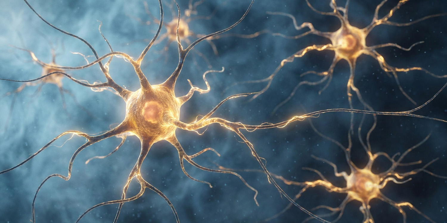
Using miniature microscopes mounted to the heads of 15 mice to track calcium ion fluctuations—which serve as a proxy for neural activity—the researchers confirmed that Drd2-positive neurons sustained elevated activation levels continuously throughout the duration of threatening auditory cues.
Proving Necessity: Silencing the Alarm
Establishing that a brain region can trigger fear is only half the scientific battle; researchers must also demonstrate that the region is strictly necessary for natural fear responses to occur.
To test this, the research team utilized inhibitory optogenetic tools to silence Drd2-positive ASt neurons in a cohort of 21 mice during threat-conditioning tasks. The results were striking. When these specific neurons were artificially silenced, the animals exhibited a nearly 50 percent reduction in natural defensive behaviors, such as freezing or rapid darting, when exposed to the threat-predicting tone.
Crucially, silencing these cells did not impair the mice’s ability to process reward cues, nor did it disrupt their general motor function or exploratory behavior in open-field environments. This selective impairment confirmed that the ASt is uniquely dedicated to orchestrating defensive survival mechanisms without compromising general cognition or baseline mobility.
"The neural and behavioral results that we observed were striking," lead author Fergil Mills noted. "Inhibiting a subpopulation of ASt neurons caused a nearly 50% reduction in fear responses—a major decrease for a structure never before established as part of the fear circuit. This tells us that the ASt is not just correlated with fear responses, but makes a meaningful causal contribution to them."
Specialized Processing of Sensory Cues vs. Environmental Context
An intriguing nuance uncovered during the study involved the specificity of ASt activation. While the region maintained sustained responses to explicit auditory threat cues, the mice did not display ASt activation when placed into spatial environments or contexts previously associated with trauma or fear.
"An interesting detail of our study was that the ASt maintains responses to specific threat cues, but not to locations where negative experiences occurred," Mills explained. "This suggests that the ASt has a specialized role in fear triggered by sensory cues, rather than the broader environment we are in—a key new insight into how the brain organizes different kinds of fear responses."
This distinction indicates that while structures like the hippocampus may encode contextual, place-based fear, the ASt operates as a dedicated sensory-cue alarm system. The researchers emphasize that these findings do not displace established models of amygdala function; rather, they position the ASt as an essential downstream partner that works in concert with the amygdala to construct a comprehensive behavioral defense strategy.
Broader Implications for Mental Health and Human Neuropsychiatry
While the empirical phase of the research was conducted in murine models, the translational implications for human health are profound. Maladaptive, persistent, and generalized fear responses represent the core diagnostic hallmarks of severe neuropsychiatric disorders, including post-traumatic stress disorder (PTSD), generalized anxiety disorder, and various phobias. In clinical populations, neural circuits that process threat detection fail to shut down appropriately, leaving patients trapped in chronic states of hyperarousal and distress.
"Understanding the circuits for fear learning is critical to human health, because maladaptive fear responses are a hallmark of neuropsychiatric disorders such as PTSD and anxiety," Mills emphasized. "In these disorders, fear responses can become persistent, generalized, or spontaneous, leading to debilitating health outcomes. In order to develop new treatments for these disorders, we have to understand the fundamental circuitry that the brain uses for fear learning. That’s what makes the discovery of the ASt’s role in fear so exciting—it’s part of a larger puzzle that we need to solve to improve human health."
Because the ASt has historically been overlooked in mental health research, its identification opens entirely new avenues for targeted pharmaceutical and neuromodulatory interventions. If ASt circuit dysfunction underpins human conditions characterized by pathological sustained fear, future therapies could potentially focus on modulating Drd2-positive signaling pathways within this transition zone.
Future Directions and Collaborative Horizons
Recognizing the limitations inherent in animal models, the research team is already taking steps to bridge the gap between basic neuroscience and clinical application. Collaborating with independent research groups, the investigators are actively initiating studies to investigate the functional role of the ASt in human subjects.
These upcoming translational efforts aim to map homologous structures in the human brain, examining whether variations in ASt volume, connectivity, or dopamine receptor density correlate with vulnerability to anxiety disorders and trauma-related pathology. Additionally, future laboratory studies will seek to untangle the precise synaptic mechanisms through which the amygdala communicates with the ASt during the initial acquisition of a threat memory.
The study, titled "Amygdalostriatal transition zone neurons encode sustained cue responses to guide defensive behaviors," was authored by Fergil Mills, Christopher R. Lee, James R. Howe, Hao Li, Maria N. Keisler, Shan Shao, Felix H. Taschbach, Mackenzie E. Lemieux, Faith Aloboudi, Jesse White, May G. Chan, Matilde Borio, Laurel R. Keyes, Hannah S. Chen, Fabiha Bushra, Gates P. Schneider, Dani P. Lemmon, Kyung J. Lee, Alexa L. Gross, Kanha Batra, Reesha R. Patel, Meenakshi M. Asokan, Jeremy Delahanty, Christian Cazares, Christopher R. Heyman, Nicholas B. Poll, Liezl Maree, Romy Wichmann, Talmo D. Pereira, Marcus K. Benna, Cory M. Root, and Kay M. Tye. Funding and institutional support were provided by the Salk Institute for Biological Studies and the University of Utah, paving the way for a new era in the neurobiology of emotion and survival.