Kang Muslim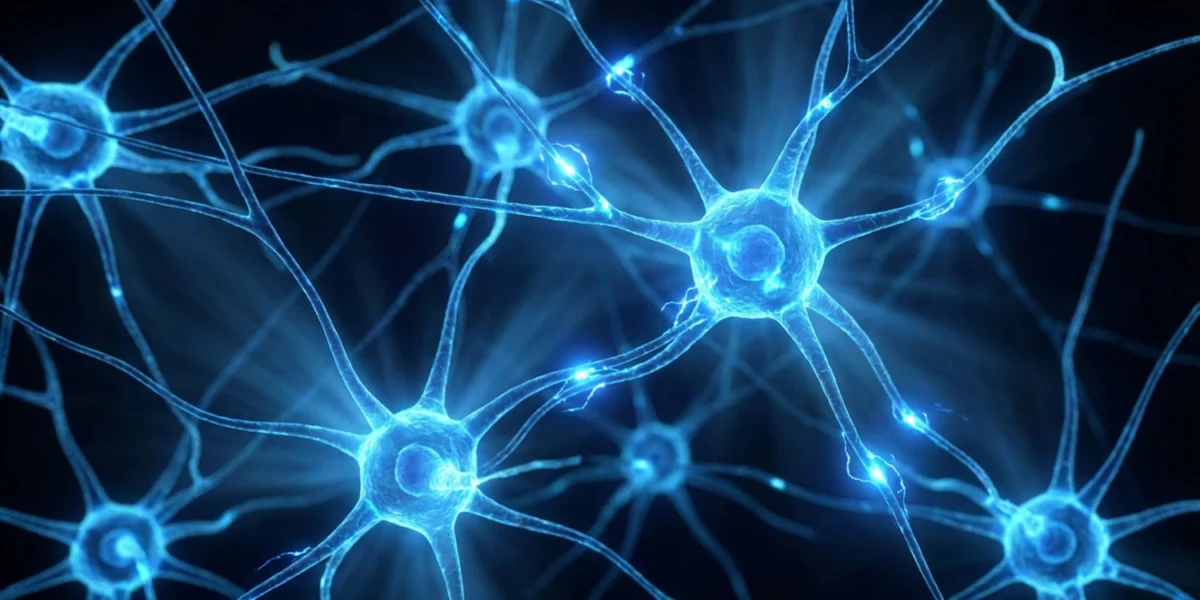
For centuries, ancient wellness traditions, contemplative practices, and modern clinical psychotherapies have prescribed slow, rhythmic nasal breathing as a primary defense against acute stress and chronic anxiety. While generations of practitioners have experienced the calming physiological effects of deep breathing exercises, the exact neurobiological mechanisms underlying this phenomenon have long remained elusive. Neuroscientists have historically debated whether breathing-induced calm is mediated entirely through top-down cognitive processes originating in the cerebral cortex, regulated automatically by respiratory centers in the lower brainstem, or driven directly by the physical sensation of airflow itself.
Recent empirical research published in the Proceedings of the National Academy of Sciences (PNAS) has decisively settled this debate. Investigators at the Institutes of Brain Science at Fudan University have mapped a direct, causal pathway connecting the mechanical frequency of airflow in the nasal cavity straight to the brain’s core emotional centers. The findings demonstrate that the sheer speed of nasal breathing acts as an independent biological signal, with slow nasal breathing actively suppressing anxiety pathways and rapid breathing accelerating them. This discovery provides rigorous empirical grounding for breathing techniques while offering a sobering physiological caveat: during periods of stress, rapid or erratic mouth breathing does not merely reflect an anxious state—it actively reinforces and exacerbates it.
The Mechanistic Mystery: Untangling Breath and Emotion
To understand how respiration shapes affective states, researchers must look beyond the mechanics of oxygen exchange and carbon dioxide regulation. As air moves through the nasal cavity during inhalation, its physical pressure and chemical composition stimulate olfactory sensory neurons (OSNs). These specialized peripheral nerve cells, nestled deep within the nasal passages, are uniquely equipped to detect both chemical odorants and the mechanical forces generated by airflow.
Previous scientific inquiries hinted at a profound structural link between respiration and neurological function. Notably, a foundational 2016 study covered by PsyPost demonstrated that the route and rhythm of breathing directly modulate cognitive processing, revealing that nasal inhalation accelerates the human brain’s ability to recognize fearful facial expressions compared to mouth inhalation. Subsequent research published in 2025 further revealed that exposure to anxiety-inducing environments naturally prompts animals to shift toward rapid breathing patterns, which in turn drives distinct shifts in neural oscillation frequencies.
Despite these clues, the precise neurocircuitry linking nasal airflow speed to emotional regulation remained poorly defined. Lead investigators sought to determine whether the mechanical frequency of the breath alone could dictate behavioral outcomes, independent of conscious psychological effort or metabolic changes in blood-gas chemistry.
Experimental Design and Optogenetic Mapping
To test the hypothesis that airflow frequency functions as an independent neural signal, a research team led by Xinsong Guo and supervised by Professor Ruiqi Wu at Fudan University designed a rigorous behavioral and neurophysiological study utilizing murine models. The researchers first established baseline respiratory parameters in mice, categorizing two breaths per second as a slow frequency, four breaths per second as a moderate frequency, and seven breaths per second as a fast frequency.
To isolate the mechanical effects of nasal airflow from actual breathing patterns, the team employed optogenetics—an advanced technique enabling precise control of neural activity using light. By genetically modifying the olfactory sensory neurons of the mice to express light-sensitive proteins, researchers implanted tiny fiber optic cables directly into the animals’ nasal cavities. This technological innovation allowed scientists to dictate the exact firing frequency of the nasal neurons using a blue laser, leaving the animals’ natural, spontaneous breathing rates entirely unaffected.
The subjects were subsequently introduced to standardized behavioral paradigms designed to measure anxiety-like states, including the Elevated Plus Maze and the Open Field Test. Because mice are evolutionarily conditioned prey animals, they exhibit an innate aversion to open, brightly lit spaces where they are vulnerable to predation. Consequently, baseline anxious rodents tend to hug the perimeter of an open box or remain restricted to the enclosed arms of a maze. Conversely, increased exploration of open, exposed areas serves as a reliable behavioral marker for reduced anxiety.
Behavioral Observations and Circuitry Identification
The behavioral data yielded clear, reproducible divisions based entirely on the frequency of optogenetic stimulation. When the nasal neurons were stimulated at a slow rate of two Hertz—simulating slow breathing—the mice exhibited a marked decrease in anxiety-related behaviors, spending significantly more time exploring the open arms of the maze and the center of the open field. When the stimulation frequency was increased to seven Hertz, mimicking rapid, panicked breathing, the subjects consistently avoided open spaces, displaying behaviors indicative of heightened anxiety. Moderate stimulation at four Hertz produced no statistically significant deviation from baseline behavioral norms.
To trace how these peripheral signals translated into emotional shifts, the research team recorded real-time electrical activity within the perirhinal cortex, a deep-brain region structurally positioned to receive direct input from the olfactory system and heavily implicated in memory and associative processing. The analysis focused specifically on high-gamma waves—rapid neural electrical oscillations associated with active information processing, cognitive engagement, and inter-regional brain communication.
The electrophysiological recordings revealed that slow nasal stimulation significantly amplified high-gamma wave activity within the perirhinal cortex. Conversely, fast nasal stimulation suppressed these high-frequency rhythms. Further tracing of the neural circuitry mapped a precise anatomical pathway: electrical impulses originating in the nasal OSNs traveled to mitral cells within the olfactory bulb, the brain’s primary olfactory relay station. From there, signals projected directly to parvalbumin-positive (PV+) interneurons residing in the perirhinal cortex. These specialized cells function as local neural pacemakers, synchronizing brain wave activity before relaying the signal downstream to the basolateral amygdala, the almond-shaped limbic structure universally recognized as the master regulator of fear and emotional reactivity.
Chemogenetic Verification and Therapeutic Implications
To confirm that this specific nose-to-brain circuit causally regulates anxiety rather than merely correlating with it, the researchers utilized chemogenetics—a method involving engineered designer receptors exclusively activated by designer drugs (DREADDs). By artificially activating the PV+ interneurons in the perirhinal cortex, the team successfully replicated the anti-anxiety effects observed during slow optical stimulation, driving up high-gamma wave production and encouraging exploratory behavior in the subjects. Conversely, selectively inhibiting these cells induced immediate anxiety-like avoidance behaviors.
In the final phase of their investigation, the researchers evaluated whether targeted slow nasal stimulation could serve as a sustainable therapeutic intervention for chronic anxiety. Using mild, unpredictable foot shocks, the team induced a persistent, anxiety-like state in a cohort of mice. These chronically stressed subjects displayed depleted high-gamma oscillations and completely avoided open testing areas.
The anxious subjects were then subjected to a daily, two-hour regimen of slow-frequency stimulation—administered via both optogenetic light pulses and direct, mechanical puffs of air delivered into the nasal passages—for a duration of two consecutive weeks. Both treatment modalities successfully restored normal exploratory behavior, repaired the underlying deficits in high-gamma brain wave synchronization, and yielded lasting reductions in anxiety-like symptoms that persisted for at least one week following the cessation of the therapy.
Broader Implications and Future Clinical Horizons
While the implications of the Fudan University study are profound, researchers emphasize several critical nuances regarding the translation of these findings to human medicine. Because the study was conducted exclusively on male murine models, future investigations must incorporate female subjects to determine whether hormonal fluctuations influence the operational dynamics of the nose-to-brain anxiety circuit. Furthermore, while optogenetic and mechanical air-puff models provide exceptional experimental control, they do not perfectly replicate the complex somatosensory experience of natural human respiration.
Crucially, the biological mechanism uncovered by the study relies strictly on airflow passing through the nasal cavity. Consequently, oral respiration—mouth breathing—fails to engage this specific olfactory-limbic pathway.
As the scientific community works toward clinical trials, the study provides a vital anatomical framework for understanding why ancient contemplative breathing exercises exert such powerful effects on human neurobiology. By identifying the nose as an underappreciated, noninvasive gateway into the brain’s limbic system, this research opens new avenues for the development of targeted, protocol-based nasal neuromodulation therapies designed to treat human anxiety disorders without relying solely on pharmaceutical interventions.