MIT neuroscientists have uncovered a surprising feature of the adult brain: millions of "silent synapses," immature neuronal connections that remain inactive until needed to form new memories. This groundbreaking discovery challenges long-held assumptions about brain plasticity and offers profound insights into the mechanisms of lifelong learning and cognitive resilience.
Unveiling a Hidden Reservoir of Neural Potential
For decades, the scientific consensus held that these "silent synapses" were a relic of early brain development, crucial for the rapid acquisition of information during infancy and childhood. It was widely believed that by the time an organism reached adulthood, these connections would have matured or been pruned away. However, the recent study, published in the prestigious journal Nature, reveals that approximately 30 percent of synapses in the adult mouse brain’s cortex remain in this dormant state. This substantial reservoir of unused connections suggests a sophisticated biological mechanism for ongoing learning without compromising established neural networks.
The implications of this finding are far-reaching. Researchers propose that this hidden pool of synapses may be the key to understanding how the adult brain continues to adapt and learn throughout life, a process that previously seemed challenging to reconcile with the need to preserve existing, vital memories.
Dimitra Vardalaki, an MIT graduate student and the lead author of the study, explained the significance of these dormant connections. "These silent synapses are actively seeking out new connections," Vardalaki stated. "When significant new information is presented, connections between the relevant neurons are selectively strengthened. This enables the brain to forge new memories without overwriting the crucial information stored in mature synapses, which are inherently more resistant to change."
The research was spearheaded by Mark Harnett, an associate professor of brain and cognitive sciences at MIT, who served as the senior author. Kwanghun Chung, an associate professor of chemical engineering at MIT, also contributed significantly to the study.
A Paradigm Shift in Understanding Adult Memory Formation
The concept of silent synapses dates back several decades, primarily observed in immature nervous systems. During early development, these connections were theorized to facilitate the brain’s absorption of vast amounts of environmental data. In rodent models, it was thought that these synapses largely disappeared by around 12 days of age, a period roughly analogous to the first few months of human life.
Despite this prevailing view, a subset of neuroscientists harbored suspicions that silent synapses might persist into adulthood. Evidence hinting at their continued presence emerged from studies investigating conditions like addiction, which is fundamentally a form of maladaptive learning. These investigations suggested that silent synapses could either reappear in the adult brain or remain present throughout its lifespan.
Furthermore, theoretical work by leading neuroscientists Stefano Fusi and Larry Abbott provided a conceptual framework for the necessity of both flexible and stable synaptic connections. Their models posited that the brain requires a dual system: some connections must be readily modifiable to accommodate new learning, while others must remain robust to preserve long-term memories. This theoretical underpinning laid crucial groundwork for the experimental validation achieved by the MIT team.
An Accidental Discovery Fueled by Advanced Imaging Technology
The MIT research team’s initial objective was not to identify silent synapses. Their work was a continuation of earlier investigations into how dendrites—the treelike extensions of neurons responsible for receiving signals—process information differently based on their location.
To delve deeper into this phenomenon, the researchers employed a cutting-edge technique known as eMAP (epitope-preserving Magnified Analysis of the Proteome). This innovative method involves physically expanding brain tissue, allowing for the precise labeling and visualization of proteins at an unprecedented level of detail. The eMAP technique enabled scientists to examine neurotransmitter receptors along dendrites with remarkable clarity.
It was during this meticulous imaging process that the researchers encountered an unexpected and remarkable observation.
"The first thing we saw, which was super bizarre and we didn’t expect, was that there were filopodia everywhere," recalled Professor Harnett, describing the moment of discovery.
Filopodia are minute, finger-like protrusions that extend from dendrites. While these structures had been documented previously, their precise function remained largely enigmatic due to their minuscule size and the limitations of conventional research tools.
Filopodia: The Unmistakable Signature of Silent Synapses
Leveraging the enhanced resolution of the eMAP technique, the MIT team detected filopodia across multiple regions of the adult mouse brain, including the visual cortex. The observed density of these structures far surpassed any previous reports. Critically, the researchers found that these filopodia were rich in NMDA receptors but conspicuously lacked AMPA receptors.
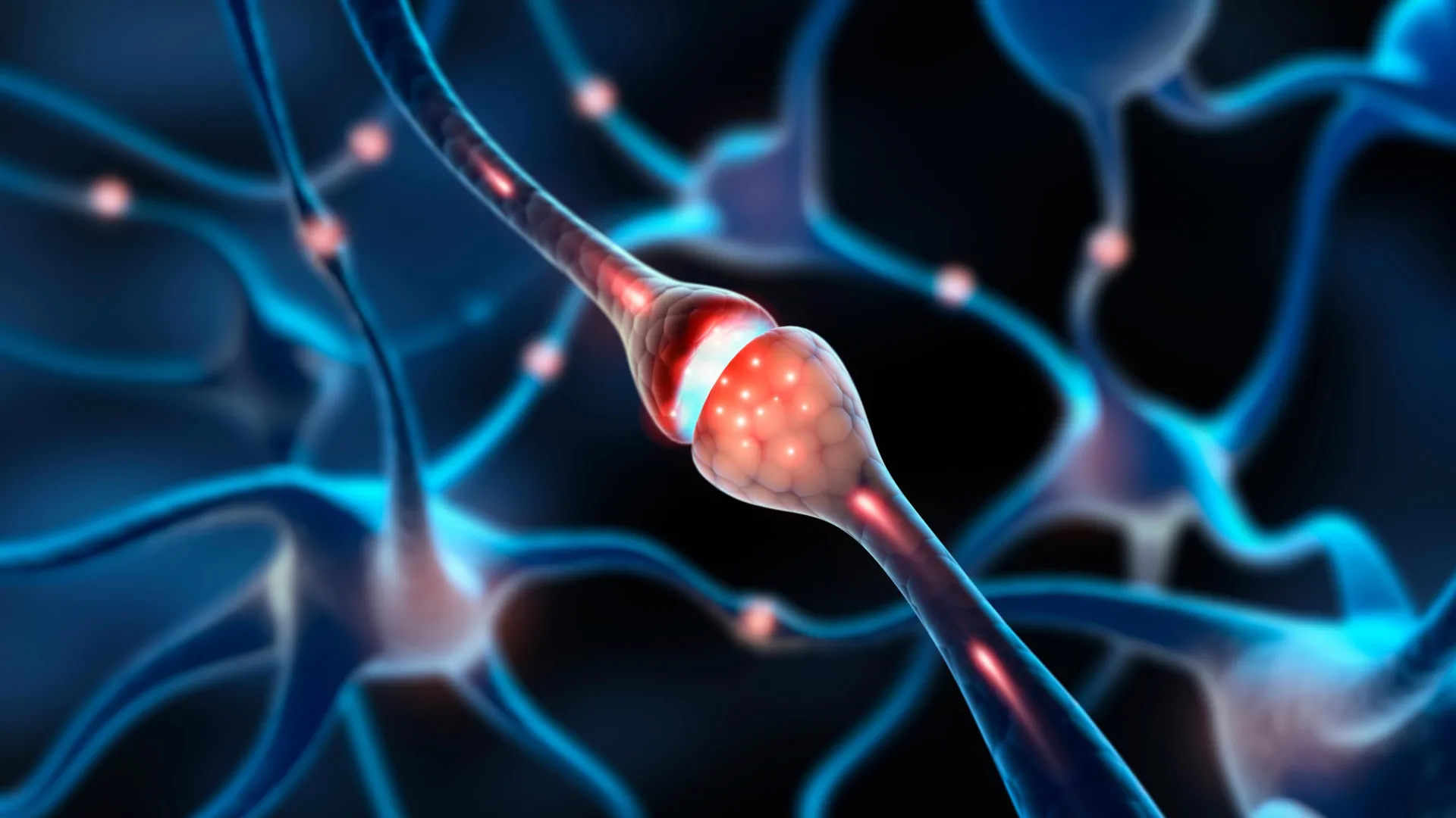
This specific molecular composition is a pivotal clue. Active synapses, the conduits for neuronal communication, typically possess both NMDA and AMPA receptors. These receptors work in concert to transmit signals using the neurotransmitter glutamate. Under normal physiological conditions, NMDA receptors alone are insufficient to pass electrical signals because they are blocked by magnesium ions. The absence of AMPA receptors in filopodia renders these connections electrically inert, hence their designation as "silent."
Activating the Dormant: The "Unsilencing" of Synapses
To rigorously test the hypothesis that these filopodia indeed function as silent synapses, the researchers employed a modified patch-clamping technique. This advanced methodology allowed them to measure electrical activity at individual filopodia while simultaneously simulating the release of glutamate, the brain’s primary excitatory neurotransmitter.
Their experiments revealed that glutamate release, in isolation, failed to elicit a discernible electrical signal. However, upon experimental unblocking of the NMDA receptors, a signal was generated. This crucial finding provided compelling evidence that these filopodial structures behave precisely as silent synapses.
Further experimentation demonstrated the remarkable plasticity of these dormant connections. The team successfully showed that it is possible to activate, or "unsilence," these synapses. By pairing glutamate release with an electrical signal from the neuron, they induced the accumulation of AMPA receptors at the synapse. This process effectively transformed the silent connection into a fully functional synapse capable of transmitting signals.
A key finding was the comparative ease with which these silent synapses could be activated. The process required significantly less stimulation compared to modifying already active synapses.
"If you start with an already functional synapse, that plasticity protocol doesn’t work," Professor Harnett elaborated. "The synapses in the adult brain have a much higher threshold, presumably because you want those memories to be pretty resilient. You don’t want them constantly being overwritten. Filopodia, on the other hand, can be captured to form new memories."
A Brain Capable of Both Flexibility and Enduring Stability
These findings offer robust support for the concept that the adult brain achieves a delicate balance between adaptability and stability by maintaining a reserve of highly malleable synapses. This dual capacity allows for continuous learning while preserving the integrity of long-term memory.
"This paper is, as far as I know, the first real evidence that this is how it actually works in a mammalian brain," Professor Harnett stated. "Filopodia allow a memory system to be both flexible and robust. You need flexibility to acquire new information, but you also need stability to retain the important information."
Implications for Aging, Neurological Health, and Lifelong Cognition
The research team is now actively pursuing the existence of similar silent synapses in human brains. A critical next step involves understanding how the prevalence and function of these connections might change with age or in the context of various neurological conditions.
"It’s entirely possible that by changing the amount of flexibility you’ve got in a memory system, it could become much harder to change your behaviors and habits or incorporate new information," Professor Harnett posited. "You could also imagine finding some of the molecular players that are involved in filopodia and trying to manipulate some of those things to try to restore flexible memory as we age."
Emerging neuroscience research continues to explore the intricate ways synaptic plasticity underpins lifelong learning. Studies focusing on the aging brain suggest that a decline in synaptic flexibility may contribute to age-related memory impairment. Concurrently, investigations into neurodegenerative diseases like Alzheimer’s highlight disruptions in synapse formation and function as key pathological features. There is a burgeoning interest in targeting these synaptic mechanisms to enhance cognitive resilience and bolster learning capacity in later life.
Collectively, these discoveries are painting an increasingly dynamic picture of the brain, one that is far more adaptable and responsive than previously imagined. Rather than being a static organ, the brain appears to maintain a hidden reserve of connections, poised for activation when new experiences and learning opportunities arise.
The research was generously supported by funding from the Boehringer Ingelheim Fonds, the National Institutes of Health, the James W. and Patricia T. Poitras Fund at MIT, a Klingenstein-Simons Fellowship, a Vallee Foundation Scholarship, and a McKnight Scholarship. This multifaceted support underscores the broad scientific community’s recognition of the profound implications of this discovery for our understanding of the brain.