Johny Marice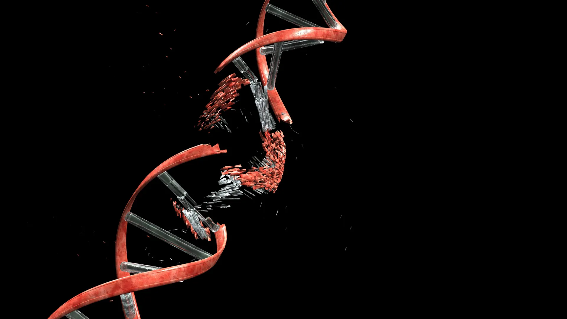
As the intricate architecture of the brain takes shape, a fundamental yet previously underappreciated challenge faces developing neurons: their arduous journey to their designated locations within the cerebral cortex. This critical phase of neurodevelopment, where newly born nerve cells must navigate a densely packed and highly organized environment, forces them through microscopic interstices, squeezing between fibrous structures and their cellular neighbors. A groundbreaking study, recently published in the prestigious journal Nature, has illuminated an astonishing consequence of this cellular odyssey: migrating neurons routinely experience substantial DNA damage, specifically double-strand breaks—a severe form of DNA injury where both strands of the DNA helix are severed.
Historically, double-strand breaks have been predominantly associated with detrimental outcomes, including mutations, cellular dysfunction, and programmed cell death (apoptosis). However, the findings from researchers at Kyoto University’s Institute for Integrated Cell-Material Sciences (WPI-iCeMS), in collaboration with several other esteemed institutions, reveal that this form of DNA damage is not an anomaly but rather an intrinsic and normal component of healthy brain cortex development. Crucially, the study demonstrates that in developing brains, these breaks are efficiently and rapidly repaired, preventing any lasting detrimental effects on the neurons or the overall neural circuitry.
"The developing brain appears to have evolved to tolerate and repair the neuronal damage efficiently," stated Professor Mineko Kengaku, the lead investigator of the study and a prominent figure at WPI-iCeMS. "But understanding the limits of that tolerance—and what happens when repair is incomplete—brings us closer to understanding a range of neurological conditions." This pivotal insight suggests that the inherent resilience of developing neurons to DNA damage is a testament to sophisticated evolutionary adaptations, and disruptions to this repair machinery could hold the key to unraveling the etiology of various neurological disorders.
The Mechanical Stress of Neuronal Migration and DNA Double-Strand Breaks
To meticulously investigate the origins and mechanisms of this DNA damage, the research team devised an ingenious experimental approach. They meticulously recreated the physical constraints and mechanical stresses encountered by migrating neurons by guiding developing neuronal cells through precisely engineered microchannels. These microchannels were designed to accurately mimic the confined and restrictive spaces that characterize the rapidly growing brain tissue during embryonic and fetal development.
Employing advanced live-cell imaging techniques and fluorescently labeled molecular probes, the scientists were able to observe in real-time the emergence of double-strand DNA breaks as the neurons navigated these narrow channels. A remarkable observation was that once the neurons successfully traversed these constricted pathways and emerged into less confined environments, the DNA damage progressively diminished. The vast majority of these double-strand breaks were found to be repaired within a 24-hour period, allowing the neurons to continue their migratory journey and subsequently integrate into the neural network without any apparent functional impairment. This rapid and effective repair mechanism underscores the brain’s remarkable capacity for self-correction during its formative stages.
The study pinpointed a key molecular player in this process: Topoisomerase IIα (also known as Top2A). This enzyme is a critical component of cellular machinery, normally tasked with managing the topological stress that naturally arises within DNA. Under physiological conditions, Top2A functions by transiently cleaving one or both DNA strands to alleviate the supercoiling and torsional tension generated by essential cellular activities, such as DNA replication and transcription. Following the relaxation of tension, the enzyme then meticulously re-ligates (reconnects) the severed DNA strands. This dynamic process can be conceptually likened to untangling a tightly wound cable by temporarily cutting it, releasing the twists, and then expertly splicing it back together.
However, the research revealed that when neurons are subjected to significant mechanical stress—such as the forces exerted during their arduous passage through narrow intercellular spaces—Top2A can become trapped in a state of incomplete cleavage. This predicament leaves sections of the DNA helix severed. In such instances, the cell must then rely on a highly conserved DNA repair pathway known as non-homologous end joining (NHEJ) to accurately re-ligate the broken DNA ends and restore the integrity of the genome. The study’s findings suggest that the mechanical forces encountered during migration are sufficient to disrupt the normal catalytic cycle of Top2A, leading to its transient malfunction and subsequent DNA breaks.
Differential DNA Damage Response: Neurons Versus Cancer Cells
A significant aspect of the study involved comparing the DNA damage response of developing neurons to that of other cell types, particularly cancer cells, which are also known for their migratory capabilities, albeit in a pathological context. The researchers found a striking difference in how neurons and cancer cells handled DNA damage when subjected to similar microchannel confinement.
In cancer cells, DNA damage often occurs in a more random and widespread fashion. This indiscriminate damage can have profound disruptive effects on critical cellular functions, leading to genomic instability, oncogenic mutations, and, ultimately, triggering programmed cell death. The uncontrolled proliferation and invasive nature of cancer cells are frequently linked to their compromised DNA repair mechanisms and their ability to tolerate higher levels of genomic insults.
In stark contrast, the DNA double-strand breaks observed in migrating neurons were not random. Instead, they were found to be predominantly localized to specific regions of the genome that are not actively transcribed or are less critical for immediate gene function. This genomic compartmentalization of damage is a crucial finding, as it suggests that essential genes, those vital for neuronal survival and function, are largely spared from severe injury. Consequently, despite the temporary presence of DNA breaks, the neurons are able to maintain their normal cellular activities and continue their developmental trajectory. This targeted nature of the damage, coupled with efficient repair, explains why neurons can withstand such significant mechanical challenges without succumbing to widespread cellular dysfunction or death.
The Clinical Implications of Impaired DNA Repair
To delve deeper into the consequences of compromised DNA repair during neuronal development, the research team ingeniously engineered mice in which newly formed cerebellar neurons were genetically modified to lack Ligase 4 (LIG4). Ligase 4 is a cornerstone enzyme of the NHEJ pathway, indispensable for the efficient repair of DNA double-strand breaks.
The initial developmental stages of these genetically engineered mice appeared outwardly normal, with no discernible abnormalities evident in their early lives. However, as these mice progressed into adulthood, they began to exhibit subtle yet progressively worsening motor coordination deficits, particularly issues related to balance. These observable symptoms bore a striking resemblance to the clinical manifestations of certain human disorders characterized by genome instability and affecting the cerebellum, a brain region crucial for motor control and coordination. This experimental model provides compelling evidence that even minor failures in the DNA repair machinery during critical developmental windows can have significant long-term neurological consequences.
The findings from this study have profound implications for our understanding of brain biology and the pathogenesis of neurological diseases. They strongly suggest that DNA breakage and subsequent repair are not merely passive responses to cellular stress but may actively participate in shaping neural circuitry and contributing to individual differences in brain function. The researchers are now keen to explore whether these early-life DNA alterations, arising from the mechanical stresses of migration and their repair, might contribute to the subtle variations observed between individual neurons. Furthermore, they aim to investigate whether dysregulation of this DNA damage and repair process could play a role in the onset or progression of neurodevelopmental disorders, such as autism spectrum disorder or intellectual disability, and neurodegenerative diseases, like Alzheimer’s or Parkinson’s disease, later in life.
"It shifts how we think about the neuronal genome," Professor Kengaku emphasized. "All neurons originate from the same DNA, but DNA damage and repair can introduce small genetic differences between individual neurons through a small mechanical journey. Some of that history may be written into the genome itself." This perspective opens up exciting new avenues of research, suggesting that the very journey of neuronal development leaves an indelible mark on the genome, potentially contributing to the remarkable diversity of neural circuits and offering new insights into the complex interplay between genetic predisposition and environmental factors in brain health and disease.
This comprehensive study, a testament to international scientific collaboration, involved a multidisciplinary team from Kyoto University, the University of Tokyo, the University of Osaka, the National University of Singapore, and the Tokyo Metropolitan Institute of Medical Science. Their collective efforts have significantly advanced our understanding of a fundamental biological process, bridging the gap between cellular mechanics, molecular repair pathways, and the intricate development of the human brain. The discovery that DNA damage, when efficiently managed, is a normal part of brain formation offers a novel lens through which to view brain development and provides crucial clues for future research into preventing and treating debilitating neurological conditions.