Kang Muslim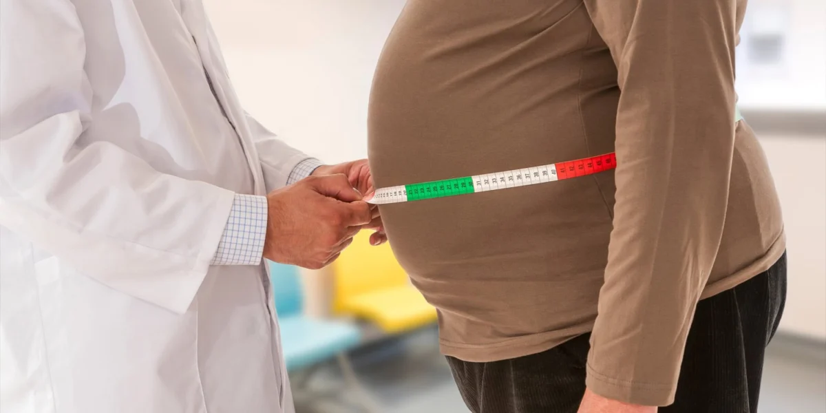
This landmark conclusion, derived from a comprehensive study published in the journal Nature Mental Health, challenges decades of clinical reliance on Body Mass Index (BMI) as the primary indicator of obesity-related health risks. Led by Professor Anqi Qiu and her research team at The Hong Kong Polytechnic University, the investigation reveals that the specific anatomical distribution of adipose tissue—where fat is stored on the body—is a more precise predictor of brain structure, neurological function, and cognitive decline than total body mass alone. By analyzing the complex relationship between regional fat depots and the aging brain, the study provides a new framework for understanding how metabolic health influences neurodegeneration in middle-aged and older populations.
For over a century, BMI has served as the global standard for assessing whether an individual is underweight, healthy, overweight, or obese. However, as a simple ratio of weight to height, BMI is notoriously blind to body composition. It cannot distinguish between muscle mass and fat, nor can it identify where fat is concentrated. The research team’s findings suggest that this "one-size-fits-all" metric may be obscuring critical insights into brain health. Specifically, the study highlights that fat stored deep within the abdominal cavity—known as visceral fat—behaves as a distinct, biologically active organ that secretes inflammatory markers, whereas fat in the limbs may have entirely different, and sometimes less deleterious, neurological associations.
The Evolution of Obesity Metrics: From BMI to Regional Adiposity
To understand the significance of this study, one must look at the chronology of how medical science has measured body fat. The Body Mass Index was developed in the mid-19th century by Adolphe Quetelet, a Belgian polymath, primarily as a tool for population statistics rather than individual clinical diagnosis. Throughout the 20th century, it became the "gold standard" due to its ease of use in clinical settings. However, by the early 2000s, the "obesity paradox" began to emerge in medical literature, where some overweight individuals appeared to have better cardiovascular outcomes than their leaner counterparts.
This paradox led researchers to investigate the "metabolically healthy obese" and the "metabolically unhealthy lean," shifting focus toward fat distribution. The emergence of advanced imaging technologies, such as Dual-energy X-ray Absorptiometry (DXA) and Magnetic Resonance Imaging (MRI), allowed scientists to move beyond the scale. The Hong Kong Polytechnic University study represents a culmination of this shift, applying these high-precision tools to a massive cohort to map the "geography of fat" against the "geography of the brain."
Methodology and the UK Biobank Dataset
The robustness of the study’s findings is rooted in its use of the UK Biobank, one of the world’s most comprehensive long-term health resources. The research team analyzed data from 18,671 participants with a mean age of approximately 62.5 years. The gender distribution was relatively balanced, with approximately 45% of the cohort being male.
The methodology was multi-layered, involving three distinct types of data collection:
- Adiposity Measurement: Instead of relying on self-reported weight or simple waist measurements, the researchers utilized DXA scans. This technology allowed them to quantify fat in four specific regions: the arms, the legs, the trunk (the central torso), and visceral fat (the deep internal fat surrounding organs).
- Neuroimaging: Participants underwent sophisticated MRI protocols. Structural MRI was used to measure the volume and thickness of brain regions; resting-state functional MRI (fMRI) tracked the communication between different neural networks; and diffusion-weighted imaging (DWI) assessed the integrity of white matter tracts—the "wiring" of the brain.
- Cognitive Assessment: A battery of tests was administered to evaluate various domains of intelligence, including fluid intelligence (reasoning and logic), prospective memory, executive function, and processing speed.
To ensure that the results reflected the impact of where fat was located rather than just how much fat was present, the researchers employed rigorous statistical controls. They mathematically isolated the effects of each fat depot, essentially "removing" the influence of total BMI to see the unique contribution of arm fat versus visceral fat, for example.
The Geography of Neurodegeneration: Regional Findings
The study’s results indicate that different fat depots are "wired" to different neurological outcomes. The findings suggest a map of influence that varies significantly across the body.
Visceral Fat: The Hidden Architect of Brain Aging
Visceral fat emerged as the most significant threat to brain health. Unlike subcutaneous fat (the fat found just under the skin), visceral fat is metabolically hyperactive. The study found a powerful correlation between high levels of visceral fat and the deterioration of the brain’s white matter. Specifically, high visceral adiposity was linked to reduced nerve fiber density and increased fluid accumulation in brain tissue, suggesting a state of chronic, low-grade neuroinflammation. This damage to the brain’s "cables" results in a breakdown of communication between distant brain regions, which manifested as lower scores across all cognitive domains tested.
Arm and Trunk Fat: Impacts on Movement and Memory
Higher concentrations of fat in the arms and the trunk were uniquely associated with the thinning of the sensorimotor cortex. This region of the brain is responsible for processing sensory input and coordinating motor output. Furthermore, arm fat showed a consistent negative association with the volume of the hippocampus. Since the hippocampus is the primary center for memory formation and spatial navigation, this link provides a potential explanation for why upper-body obesity is often a precursor to memory-related disorders in later life.
Leg Fat and the Limbic System
In a surprising turn, leg fat showed a distinct relationship with the limbic system, the brain’s emotional and reward center. Higher leg fat was associated with weakened functional connectivity within this system. The researchers hypothesized that this might be related to leptin, a hormone produced by fat cells that regulates hunger and energy balance. Lower-body fat tends to secrete higher levels of leptin than upper-body fat. While leptin is necessary for health, chronic "leptin resistance"—often found in those with high adiposity—can interfere with the limbic system’s ability to regulate mood and memory.
The "Brain Age" Phenomenon
A critical component of the study was the calculation of "Brain Age." Using computer modeling and machine learning, the researchers compared the participants’ actual chronological age with the "age" suggested by their neural networks.
The data revealed that regional fat accelerates the aging process of specific brain networks, most notably the sensorimotor network, the limbic network, and the default mode network (which is active during internal thought and self-reflection). The researchers found that this "accelerated brain aging" was the primary pathway through which fat distribution harmed cognitive performance. In essence, high levels of visceral and trunk fat do not just cause immediate cognitive slips; they essentially "fast-forward" the brain’s biological clock, making a 60-year-old brain function like that of a 70-year-old.
Biological Mechanisms: Why Location Matters
The study suggests several biological reasons why fat distribution is so influential. Adipose tissue is not merely a storage site for excess calories; it is an active endocrine organ.
- Inflammatory Cascades: Visceral fat is known to secrete high levels of pro-inflammatory cytokines, such as Interleukin-6 (IL-6) and Tumor Necrosis Factor-alpha (TNF-α). These chemicals can cross the blood-brain barrier, triggering microglial activation—the brain’s immune response. When this response is chronic, it leads to the destruction of healthy neurons and synapses.
- Metabolic Signaling: Different fat depots have different sensitivities to insulin and different rates of lipolysis (the breakdown of fats). The trunk and visceral regions are more prone to releasing free fatty acids into the bloodstream, which can lead to systemic insulin resistance, a condition often referred to as "Type 3 Diabetes" when it specifically affects the brain.
- Hormonal Variance: The secretion of adiponectin, a hormone that protects the brain and improves insulin sensitivity, is often higher in leg fat and lower in visceral fat. This may explain why leg fat, in some previous studies, appeared to be less damaging or even protective compared to abdominal fat.
Implications for Public Health and Clinical Practice
The implications of this research for the medical community are profound. If BMI is an insufficient metric, healthcare providers may need to adopt more nuanced tools for assessing risk.
1. Beyond the Scale: Clinicians may need to place greater emphasis on waist-to-hip ratios or waist-to-height ratios, which are better proxies for visceral fat than BMI. In high-risk cases, the use of DXA scans—traditionally used for bone density—may become a more common tool for body composition analysis.
2. Personalized Weight Loss: The study suggests that "not all weight loss is created equal." Targeted interventions aimed at reducing visceral fat, such as high-intensity interval training (HIIT) or specific dietary adjustments (like reducing processed sugars which contribute to liver and visceral fat), may be more effective for preserving cognitive function than general calorie restriction.
3. Early Screening for Cognitive Decline: By identifying individuals with high visceral adiposity in their 40s and 50s, doctors may be able to intervene decades before the onset of symptoms like dementia or Alzheimer’s disease.
Limitations and the Path Forward
While the study is one of the largest of its kind, the authors noted several limitations that provide a roadmap for future research. The cross-sectional nature of the data means that while a strong association exists, the study cannot definitively prove that fat causes brain changes. It is possible, though less likely, that neurological changes in the brain regions governing appetite could lead to specific fat accumulation patterns.
Additionally, the UK Biobank cohort is predominantly white and British. Because body fat distribution varies significantly across different ethnic and racial groups—for example, South Asian populations tend to accumulate more visceral fat at lower BMIs than Europeans—further research is needed to see if these patterns hold true globally.
The researchers also pointed out that while DXA scans are highly accurate, they still struggle to perfectly distinguish between subcutaneous trunk fat and visceral trunk fat. Future studies using more granular 3D imaging could further isolate these effects.
Analysis of Broader Societal Impact
As the global population ages and obesity rates continue to rise, the intersection of metabolic health and neurodegeneration is becoming a primary concern for public health policy. The "dementia epidemic" is often discussed in isolation from the "obesity epidemic," but this study suggests they are two sides of the same coin.
If regional adiposity is a "shaper" of the brain, then urban planning (promoting walkable cities to reduce trunk fat) and food policy (taxing ingredients that contribute to visceral adiposity) are not just metabolic health initiatives—they are brain health initiatives. The work of Anqi Qiu and her colleagues reinforces the idea that the brain is not an isolated organ, but one deeply integrated into the body’s metabolic landscape.
As stated in the study’s conclusion, characterizing the associations between regional adiposity and brain health is essential for developing "targeted prevention and intervention strategies for cognitive aging." The message to the public is clear: it is not just the number on the scale that matters, but where you carry your weight that may ultimately determine the longevity of your mind.