Kang Muslim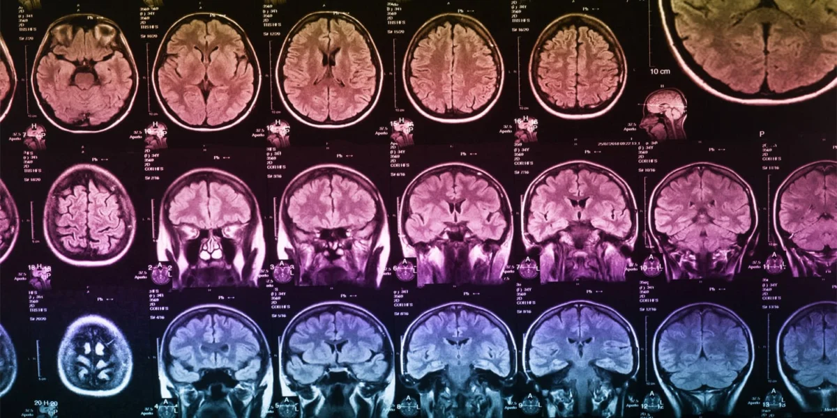
The Mental Workspace: Understanding Working Memory
Working memory functions as the brain’s "mental workspace," an essential cognitive system responsible for the temporary storage and active processing of information. Unlike long-term memory, which stores facts and experiences over years, working memory is the engine of "online" cognition. It is what allows a person to remember the beginning of a sentence while reading the end, to perform mental arithmetic, or to follow a multi-step set of directions.
This cognitive capacity is not a monolithic entity but is generally understood by psychologists to consist of several interacting components. These include a "mental sketchpad" for visual and spatial information, a "phonological loop" for auditory and verbal data, and a central executive system that coordinates attention and manages goal-directed behavior. Despite its critical importance to daily functioning, working memory is notably sensitive to the aging process. As individuals move into middle and older adulthood, the efficiency of this mental workspace typically begins to diminish, impacting everything from decision-making to the ability to learn new skills.
A Massive Dataset: The UK Biobank Advantage
To move beyond the limitations of previous small-scale studies, which often relied on narrow sample sizes or individuals with existing brain trauma, the researchers utilized data from the UK Biobank. This large-scale biomedical database and research resource contains in-depth genetic and health information from half a million UK participants. For this specific study, the researchers filtered the database to focus on a healthy cohort of 30,640 adults aged between 51 and 80.
The scale of this study is significant because it allows for high statistical power, enabling the researchers to account for numerous confounding variables that have plagued prior neurological research. By adjusting for age, biological sex, years of formal education, and total intracranial volume—the physical size of the head and brain cavity—the team could isolate the specific relationship between the volume of individual brain regions and cognitive performance with a level of precision rarely seen in neuropsychological literature.
Methodology: MRI Precision and the Numeric Memory Test
The study integrated two primary forms of data collection: advanced neuroimaging and computerized cognitive assessment. Each of the 30,640 participants underwent a magnetic resonance imaging (MRI) scan, which provides a high-resolution map of the brain’s internal structures. Using specialized automated software, the research team segmented these images to measure the volume of gray matter—the tissue containing the cell bodies of neurons—in 25 pre-selected regions of interest known to be involved in memory tasks.
Simultaneously, participants completed the Numeric Memory Test, a variation of the traditional "digit span" task. In this assessment, a sequence of numbers is displayed on a screen for three seconds before disappearing. The participant must then use a digital keypad to enter the sequence in reverse order. This "backward" requirement is a critical metric of working memory, as it forces the brain not just to store the numbers, but to actively manipulate and reorder them. As the test progresses, the sequences become increasingly longer, reaching up to 12 digits, pushing the limits of the participant’s cognitive capacity.
The Six Pillars of Working Memory
The analysis revealed that larger physical volumes in six specific brain regions were robust predictors of higher scores on the memory test. These regions span across different functional networks of the brain, suggesting that working memory relies on a highly integrated system rather than a single "memory center."
- The Cerebellum: Traditionally associated with motor control and physical coordination, the cerebellum’s role in cognitive tasks has become increasingly recognized. In this study, the researchers suggest the cerebellum assists in "inner speech"—the silent repetition of numbers that people use as a strategy to keep information active in their minds.
- The Hippocampus: Located deep within the temporal lobe, the hippocampus is the brain’s primary hub for long-term memory. However, this study reinforces the idea that it is also vital for encoding short-term visual information, acting as an entry point for the "mental workspace."
- The Superior Temporal Cortex: Situated on the sides of the brain, this region is involved in processing auditory information and language. Its involvement in a visual number test suggests that participants likely "translate" the visual digits into sounds (verbalizing them in their heads) to improve recall.
- The Insula: This region plays a critical role in the "salience network," which helps the brain switch between different mental tasks and ignore distractions. A larger insula volume likely helps individuals maintain focus on the number sequence while blocking out external or internal interruptions.
- The Left Inferior Parietal Cortex: This area is a key node for spatial processing and attention. In the context of the digit span task, it likely functions as the "mental chalkboard," allowing the individual to visualize the numbers and rearrange them in reverse order.
- The Left Lateral Occipital Cortex: Primarily involved in visual object recognition, this region assists in the initial perception and maintenance of the visual representation of the digits.
The "Education Buffer" and Demographic Influences
Beyond the physical structure of the brain, the study highlighted the profound impact of demographic factors on cognitive health. One of the most striking findings was the "protective role" of education. Participants with more years of formal schooling consistently outperformed those with less education, even when their brain volumes were similar. This supports the "cognitive reserve" hypothesis—the idea that mental stimulation and education build a more resilient brain that can function efficiently despite the natural physical changes associated with aging.
Conversely, age remained the strongest predictor of decline. As participants moved from their 50s into their 70s and 80s, the average number of digits they could successfully reverse decreased. Interestingly, the study found no significant difference in performance based on biological sex, nor did the overall size of the head predict memory capacity, suggesting that the integrity and volume of specific regions are far more important than the overall size of the organ.
Complexity in Connectivity: The Negative Correlations
In a surprising twist, the researchers found that after accounting for all variables, three specific brain regions showed a negative relationship with memory performance—meaning that in those areas, smaller volume was associated with better scores. Sarah Ellen Carnemolla noted that this likely reflects the brain’s "highly interconnected nature."
In neurological terms, more volume is not always better; sometimes, a more "streamlined" or "efficiently pruned" neural pathway can lead to faster processing. This finding underscores the complexity of the human connectome and suggests that the relationship between brain size and intelligence is not purely linear, but is influenced by how different regions communicate and share the cognitive load.
Clinical Benchmarks and Future Directions
One of the most immediate practical applications of this research is the creation of benchmark scoring tables. By using the data from 30,000 healthy individuals, the researchers have established a "gold standard" for what constitutes normal memory performance across different age groups and education levels. Doctors and clinicians can now use these tables to evaluate patients who express concerns about their memory. If a patient’s score falls significantly below the benchmark for their specific demographic, it could serve as an early warning sign for neurodegenerative conditions like dementia or Alzheimer’s disease.
However, the study authors are careful to note the limitations of their work. Because the data is cross-sectional—taken at a single point in time—it cannot definitively prove that shrinking brain regions cause memory loss. It only establishes a strong correlation. Furthermore, the UK Biobank population is not perfectly representative of the global population, as it leans toward individuals of White ethnic backgrounds and higher socioeconomic status.
Implications for Global Health
As the world faces a "silver tsunami"—a massive increase in the elderly population—understanding the neurological basis of working memory is no longer just an academic pursuit; it is a public health necessity. Conditions like ADHD and various forms of dementia are characterized by deficits in working memory, and by identifying the specific structures that support this function, researchers can begin to develop more targeted therapies.
The study also highlights the potential of neuroplasticity—the brain’s ability to reorganize itself by forming new neural connections throughout life. If we know which regions are critical for memory, we can design cognitive exercises or lifestyle interventions aimed specifically at maintaining the "integrity" of those areas.
In the words of Carnemolla, "the health of our brain underpins many everyday abilities we often take for granted." This research serves as a vital step toward a future where cognitive decline is not seen as an inevitable part of aging, but as a manageable aspect of human health that can be supported through early detection, education, and targeted neurological care. By providing a clear map of the aging brain’s mental workspace, this study offers hope for maintaining quality of life well into the later years of the human lifespan.