
Researchers at the Massachusetts Institute of Technology (MIT) have made a groundbreaking discovery in optical physics, identifying an unexpected phenomenon that could dramatically enhance the speed and resolution of imaging living tissues. Under precise, yet achievable, conditions, a laser beam that typically scatters and disperses within an optical fiber can spontaneously reorganize itself into a remarkably narrow and highly focused “pencil beam.” This emergent property has been leveraged by the MIT team to achieve three-dimensional (3D) imaging of the human blood-brain barrier at speeds approximately 25 times faster than current gold-standard techniques, all while maintaining comparable image quality. The implications of this breakthrough extend to real-time observation of individual cells absorbing drugs, offering a significant advancement for evaluating the efficacy of treatments for debilitating neurological conditions such as Alzheimer’s disease and Amyotrophic Lateral Sclerosis (ALS).
The Unexpected Emergence of a Focused Beam
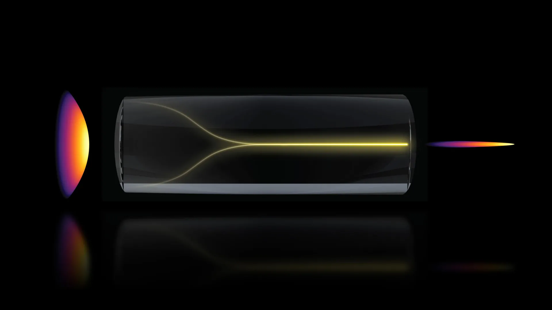
The discovery emerged from experiments conducted by Sixian You, an assistant professor in MIT’s Department of Electrical Engineering and Computer Science (EECS) and a senior author on the paper detailing the findings, and her research group. Their work began with a sophisticated device known as a “fiber shaper,” designed to meticulously control laser light as it travels through a multimode optical fiber. Multimode fibers are advantageous for their capacity to handle high power levels, a crucial factor for deep tissue penetration in imaging.
The prevailing scientific understanding in the field posited that increasing laser power within such fibers would inevitably lead to increased scattering and chaotic light behavior due to inherent imperfections within the fiber material. This chaos typically necessitates complex optical engineering to counteract, often involving custom beam-shaping components to restore focus and clarity. However, as lead author Honghao Cao, an EECS graduate student, incrementally increased the laser power to test the fiber’s limits, an anomaly occurred. Instead of the anticipated chaotic diffusion, the light unexpectedly coalesced into a single, extraordinarily sharp beam as the power approached a critical threshold, a point where the fiber itself might be susceptible to damage.
"The common belief in the field is that if you crank up the power in this type of laser, the light will inevitably become chaotic," explained Professor You. "But we proved that this is not the case. We followed the evidence, embraced the uncertainty, and found a way to let the light organize itself into a novel solution for bioimaging." This spontaneous self-organization bypasses the need for intricate external optical adjustments, offering a simpler and more robust approach to generating a high-quality beam.
Unlocking the Conditions for Self-Organization
The MIT team meticulously investigated the specific parameters that trigger this remarkable self-organizing behavior. They identified two critical requirements that, when met simultaneously, enable the transformation of a scattered laser signal into a coherent pencil beam.
The first requirement is an exceptionally precise alignment of the incoming laser beam. It must enter the optical fiber at a perfectly zero-degree angle, a level of precision significantly more stringent than typically employed in standard fiber optics applications. This meticulous alignment ensures that the light enters the fiber in a way that minimizes initial distortions and sets the stage for subsequent self-organization.
The second crucial factor involves the laser power. The power must be elevated to a point where the light begins to interact directly with the glass material of the optical fiber itself. This interaction, known as nonlinear optical effects, is key. At this critical power level, the inherent disorder within the fiber is effectively counteracted by the emergent nonlinearities.
"At this critical power, the nonlinearity can counter the intrinsic disorder, creating a balance that transforms the input beam into a self-organized pencil beam," stated Cao. This delicate balance is the cornerstone of the phenomenon. Normally, researchers tend to avoid such high power levels precisely to prevent damage to the optical fibers. Furthermore, the robustness of multimode fibers to handle substantial energy often means that extreme precision in alignment is not deemed essential for many applications. However, by deliberately combining these less-explored conditions – extreme alignment and critical power – the MIT team has demonstrated the ability to generate a stable, high-quality beam without relying on complex and expensive custom optical components.
“That is the charm of this method – you could do this with a normal, optical setup and without much domain expertise,” Professor You added, highlighting the accessibility of the technique.
Enhanced Imaging Capabilities: Speed, Detail, and Reduced Artifacts
The newly generated pencil beam exhibits exceptional stability and a remarkable level of detail when compared to conventional beams used in imaging. A common challenge with many current laser-based imaging techniques is the presence of “sidelobes” – diffuse halos surrounding the main beam that can obscure fine details and reduce overall image clarity. The self-organized pencil beam, however, remains exceptionally clean and tightly focused, significantly minimizing these artifacts and leading to sharper, more interpretable images.
To validate the practical utility of this breakthrough, the researchers applied their novel imaging technique to visualize the human blood-brain barrier (BBB). The BBB is a complex and vital cellular layer that acts as a highly selective gatekeeper, protecting the brain from harmful substances circulating in the bloodstream. However, this protective function also poses a significant challenge for the delivery of therapeutic drugs to treat neurological disorders. Many promising drug candidates are unable to penetrate this barrier, severely limiting their therapeutic potential.
Revolutionizing Drug Delivery Assessment: Faster 3D Imaging of the BBB
Understanding how drugs navigate the intricate network of blood vessels within the BBB and whether they successfully reach the brain tissue is paramount for developing effective treatments. Traditional optical methods for imaging this process typically involve capturing a series of two-dimensional (2D) slices, which are then computationally assembled to create a 3D representation. This multi-slice approach is time-consuming and can introduce cumulative errors, potentially leading to a loss of temporal resolution and detail.
The MIT team’s innovative pencil beam approach dramatically accelerates this process. By utilizing the highly focused beam, they were able to generate rapid, high-precision 3D images of the BBB. Crucially, this allowed them to track, in real-time, how cells within the barrier absorb specific proteins. This capability is particularly valuable for assessing drug efficacy.
Professor Roger Kamm, the Cecil and Ida Green Distinguished Professor of Biological and Mechanical Engineering at MIT and a co-author on the paper, emphasized the significance for the pharmaceutical industry. "The pharmaceutical industry is especially interested in using human-based models to screen for drugs that effectively cross the barrier, as animal models often fail to predict what happens in humans," Professor Kamm stated. "That this new method doesn’t require the cells to have a fluorescent tag is a game-changer. For the first time, we can now visualize the time-dependent entry of drugs into the brain and even identify the rate at which specific cell types internalize the drug." The absence of the need for fluorescent tagging simplifies experimental protocols and potentially reduces interference with drug behavior.
Sarah Spitz, a postdoc in EECS and another co-author, further elaborated on the broad applicability of the technique. "Importantly, however, this approach is not limited to the blood-brain barrier but enables time-resolved tracking of diverse compounds and molecular targets across engineered tissue models, providing a powerful tool for biological engineering," Spitz remarked. This suggests that the technology could be adapted to study drug interactions and cellular uptake in a wide array of biological contexts beyond the brain.
The results of the tests were striking: the system generated cellular-level 3D images with demonstrably improved quality, and it achieved this at a pace approximately 25 times faster than existing state-of-the-art methods. This speed improvement is critical for observing dynamic biological processes that occur over short timescales.
Professor You highlighted another key advantage: overcoming a common trade-off in microscopy. "Usually, you have a tradeoff between image resolution and depth of focus – you can only probe so far at a time. But with our method, we can overcome this tradeoff by creating a pencil-beam with both high resolution and a large depth of focus," she explained. This means that the system can capture detailed images from a greater depth within the tissue without sacrificing clarity, further enhancing its utility for studying complex biological structures.
A Timeline of Discovery and Future Directions
The journey leading to this discovery can be traced back to prior work by the MIT team, specifically their development of an advanced fiber shaper. This foundational research, conducted over an unspecified period prior to the recent findings, laid the groundwork for manipulating laser light with high precision within optical fibers.
The critical phase of the current research likely began approximately one to two years ago, with Honghao Cao’s systematic testing of the fiber shaper’s performance limits by gradually increasing laser power. This iterative process of experimentation and observation, spanning several months, led to the unexpected identification of the self-organizing beam phenomenon. Following this initial observation, the team dedicated significant effort to characterizing the precise conditions required for its reproduction and to exploring its imaging capabilities. The publication in Nature Methods today marks the culmination of this intensive research period, indicating a substantial effort in validation and manuscript preparation over the past year.
The research team, comprising Honghao Cao (lead author, EECS graduate student), Sixian You (senior author, EECS assistant professor), Li-Yu Yu (EECS graduate student), Kunzan Liu (EECS graduate student), Sarah Spitz (postdoc), Francesca Michela Pramotton (postdoc), Federico Presutti (postdoc), Zhengyu Zhang (PhD ’24), Subhash Kulkarni (assistant professor at Harvard University and the Beth Israel Deaconess Medical Center), and Roger Kamm (Cecil and Ida Green Distinguished Professor of Biological and Mechanical Engineering at MIT), represents a multidisciplinary effort drawing expertise from electrical engineering, computer science, physics, and biological and mechanical engineering.
Broader Implications and Next Steps
The potential impact of this self-organizing laser beam technology extends far beyond the immediate applications in neurological drug development. By offering a faster, more detailed, and less artifact-prone imaging modality, it could revolutionize various fields of biomedical research. This includes, but is not limited to, studying cellular dynamics in cancer research, observing the effects of therapies on infectious diseases, and advancing fundamental understanding of cellular processes in developmental biology.
Looking ahead, the MIT researchers are focused on several key areas. A primary objective is to delve deeper into the fundamental physics governing the self-organization of light within optical fibers. Understanding the precise mechanisms at play will be crucial for optimizing the technique and potentially discovering even more advanced optical phenomena.
Furthermore, the team plans to broaden the application of this imaging method to other biological systems. This includes imaging individual neurons to understand brain function and dysfunction, and adapting the technology for the study of other complex tissues and organs. The ultimate goal is to translate this laboratory breakthrough into practical, accessible tools for researchers and clinicians worldwide.
The research was supported by a consortium of funding sources, including MIT startup funds, the National Science Foundation (NSF), the Silicon Valley Community Foundation, the Diacomp Foundation, the Harvard Digestive Disease Core, a MathWorks Fellowship, and the Claude E. Shannon Award. This diverse financial backing underscores the perceived importance and broad potential of this innovative imaging technology. The successful demonstration of this novel optical phenomenon by the MIT team marks a significant step forward in our ability to visualize and understand the intricate workings of living systems.


