Johny Marice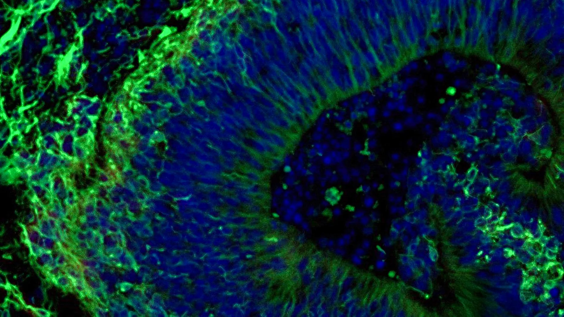
Scientists at Johns Hopkins Medicine have uncovered groundbreaking evidence suggesting that miniature clusters of brain tissue, cultivated from the cells of individuals with Alzheimer’s disease, can serve as a critical tool in predicting how patients might respond to medications aimed at managing the condition’s challenging neuropsychiatric symptoms. This pioneering research, published in Alzheimer’s & Dementia: The Journal of the Alzheimer’s Association, represents a significant stride towards developing more precise and individualized therapeutic approaches for the millions affected by this debilitating neurodegenerative disorder.
The Power of Miniature Brain Models
The core of this transformative research lies in the creation and study of laboratory-grown brain tissues, known as organoids. These three-dimensional structures, meticulously engineered to mimic aspects of the human brain, are offering an unprecedented window into the complex mechanisms of Alzheimer’s disease. By utilizing organoids derived from patient cells, researchers can observe disease-specific molecular changes in a controlled environment, a feat previously unattainable with traditional cell cultures or animal models alone.
Alzheimer’s disease, the most prevalent form of dementia, currently impacts more than 7 million Americans, a number projected to rise significantly in the coming decades due to an aging global population. While a cure remains elusive, a significant portion of patients experience neuropsychiatric symptoms such as anxiety, depression, agitation, and psychosis. These symptoms profoundly diminish quality of life for both patients and their caregivers, and current treatment strategies, often involving selective serotonin reuptake inhibitors (SSRIs), yield highly variable results. This variability underscores the urgent need for methods that can anticipate individual patient responses to medication.
The Johns Hopkins team’s work adds substantial weight to the growing body of evidence supporting the utility of these miniature brain models. Beyond predicting drug efficacy, the research also identified tiny particles, called extracellular vesicles, released by these organoids. These vesicles, acting as cellular messengers, may hold the key to developing novel biomarkers for early diagnosis and for gauging the progression of Alzheimer’s disease.
A Deeper Dive into Hindbrain Organoids and SSRI Efficacy
The study specifically focused on hindbrain organoids. The hindbrain, located at the posterior part of the brain, is crucial for regulating fundamental life functions including breathing, sleep-wake cycles, and heart rate. Understanding how Alzheimer’s disease affects this region and how medications might influence it is vital. The researchers hypothesized that these hindbrain organoids could reveal molecular signatures indicative of a patient’s potential benefit from escitalopram oxalate, a commonly prescribed SSRI used to treat depression and anxiety, and often employed to manage neuropsychiatric symptoms in Alzheimer’s patients.
The scientific journey began with the meticulous collection of blood samples, obtained with informed consent, from individuals diagnosed with Alzheimer’s disease at the National Institutes of Health (NIH)-funded Johns Hopkins Alzheimer’s Disease Research Center. This initial step was critical for establishing a direct link between patient biology and the experimental models.
From Blood Cells to Brain Tissue: The Reprogramming Process
A key innovation in the Johns Hopkins study was the sophisticated technique used to generate the brain organoids. The researchers employed a process of cellular reprogramming to revert the collected blood cells into a pluripotent stem cell-like state. These induced pluripotent stem cells (iPSCs) possess the remarkable ability to differentiate into virtually any cell type in the body.
By utilizing iPSCs derived from both individuals with Alzheimer’s disease and healthy control participants, the team was able to create hindbrain organoids. These organoids were not merely generic brain tissue; they were specifically designed to contain specialized brain cells, or neurons, that produce serotonin, a neurotransmitter heavily implicated in mood regulation and targeted by SSRIs.
The iPSCs were then carefully guided through a developmental process, encouraging them to self-organize into small, pea-sized clusters of brain tissue that closely mirrored the structural organization of the hindbrain. The scale of this undertaking was substantial; the study incorporated hundreds of organoids, each representing an individual patient with Alzheimer’s disease or a healthy participant. Dr. Vasiliki Machairaki, the study’s lead investigator and an associate professor of genetic medicine at Johns Hopkins University School of Medicine, noted that this likely represents one of the most extensive brain organoid studies conducted to date in the field of Alzheimer’s research. This large-scale approach enhances the statistical power and reliability of the findings.
Unveiling Molecular Hallmarks of Alzheimer’s in Organoids
A significant outcome of the research was the successful replication of several key biological characteristics of Alzheimer’s disease at the molecular level within the patient-derived organoids. When compared to organoids grown from the cells of healthy individuals, those originating from individuals with Alzheimer’s exhibited distinct differences in the expression of proteins involved in crucial cellular processes. These included proteins vital for communication between brain cells (synaptic function), inflammatory responses, and pathways known to be dysregulated in Alzheimer’s disease.
The subsequent phase of the study involved exposing these organoids to escitalopram oxalate. The results were illuminating. In a subset of organoids derived from Alzheimer’s patients, the medication triggered an increase in proteins associated with serotonin signaling and inter-neuronal communication. These are precisely the pathways that antidepressants are designed to modulate. However, in other organoids, the molecular response was minimal or entirely absent.
"We used these organoids to model how some patients’ tissue may respond to a commonly prescribed SSRI," explained Dr. Machairaki. "On a large-scale level, our model may eventually be used to identify subgroups of patients, based on underlying molecular mechanisms, who are more likely to respond to certain drugs and thus help us to create precise, targeted treatments in the long run." This observation directly addresses the clinical challenge of variable drug responses and points towards a future where treatment selection is guided by an individual’s unique biological profile.
Extracellular Vesicles: Potential Biomarkers for Diagnosis and Treatment Response
The researchers then turned their attention to extracellular vesicles (EVs), small membrane-bound particles that cells release into their surroundings. EVs are known to carry a cargo of proteins, lipids, and nucleic acids, effectively acting as messengers that can influence the behavior of recipient cells. The team investigated whether EVs released by the Alzheimer’s organoids could serve as biomarkers for the disease itself or, crucially, as indicators of how brain tissue responds to therapeutic interventions.
Before and after administering escitalopram treatment to the organoids, the scientists meticulously analyzed the protein content within EVs secreted by both patient-derived and healthy control organoids. The findings were compelling. The EVs were found to contain proteins integral to essential brain functions, including neuronal communication, memory formation, and neurotransmitter release.
Notably, EVs from organoids derived from individuals with Alzheimer’s disease displayed discernible alterations in several proteins linked to the disease pathology. Specifically, the levels of proteins such as RAB3A, NSF, and ATCAY were found to be reduced in the Alzheimer’s organoids. These proteins play critical roles in the normal signaling processes between neurons.
Following escitalopram treatment, a differential response was observed. In certain samples, the levels of some proteins within the EVs increased, particularly those associated with serotonin signaling and synaptic pathways that are the targets of antidepressant medications. This differential response, where some organoids exhibited a robust molecular reaction while others showed little to no change, offers significant implications.
"Some organoids displayed strong molecular responses, while others showed little or no change," Dr. Machairaki elaborated. "This variation raises the possibility that extracellular vesicles from brain organoids could eventually help identify which patients are most likely to benefit from a particular treatment." This discovery opens the door to the development of non-invasive diagnostic and prognostic tools, potentially transforming how Alzheimer’s disease is managed.
The Future of Brain Organoids: Towards a "Liquid Biopsy"
Looking ahead, Dr. Machairaki and her team are focused on enhancing the complexity and realism of their brain organoid models. Future iterations are planned to incorporate immune cells and vascular-like networks that mimic the intricate structure of blood vessels. The inclusion of these elements is expected to further bridge the gap between laboratory models and the living human brain, leading to even more accurate insights.
With continued research and refinement, Dr. Machairaki envisions a future where extracellular vesicles harvested from brain organoids could function as a form of "liquid biopsy." Such a diagnostic tool would be revolutionary, potentially enabling the early detection of Alzheimer’s disease, precise staging of its progression, and the identification of specific disease subtypes unique to each patient. This personalized approach to diagnosis could then inform tailored treatment strategies, maximizing therapeutic efficacy and minimizing adverse effects.
The current study, while a significant breakthrough, is acknowledged by the researchers as an early but vital step towards realizing this ambitious vision. The implications of this research are far-reaching, suggesting a paradigm shift in how we understand, diagnose, and treat Alzheimer’s disease, moving towards an era of truly personalized medicine.
Broader Impact and Future Directions
The implications of this research extend beyond the immediate clinical application of predicting SSRI response. The ability to generate patient-specific brain organoids that recapitulate key disease features provides an invaluable platform for drug discovery and development. Pharmaceutical companies could potentially screen novel therapeutic compounds on these organoids, identifying promising candidates and predicting their efficacy and safety profiles before costly and time-consuming human trials. This could accelerate the development of much-needed treatments for Alzheimer’s disease.
Furthermore, the identification of specific protein changes within extracellular vesicles offers a tangible pathway toward developing new diagnostic tests. Current diagnostic methods for Alzheimer’s often rely on cognitive assessments, expensive brain imaging, and invasive cerebrospinal fluid analysis. The prospect of a blood-based test, derived from EV analysis, would represent a significant advancement in accessibility and early detection, allowing for interventions at earlier stages of the disease when they are likely to be most effective.
The timeline of this research, from sample collection to publication, represents years of dedicated scientific inquiry. The initial groundwork laid by the Johns Hopkins team and their collaborators, including researchers from the University of Rochester School of Medicine and Dentistry and Tymora Analytical Operations, underscores the collaborative nature of modern scientific endeavors. Funding from prestigious institutions like the National Institutes of Health, the Paul G. Allen Frontiers Foundation, and The Johns Hopkins University’s own Precision Medicine Center highlights the recognized importance and potential of this research avenue.
While the scientific community awaits further validation and translation of these findings into clinical practice, the current study offers a powerful beacon of hope. The development of sophisticated brain organoid models, coupled with the analysis of extracellular vesicles, is paving the way for a future where Alzheimer’s disease is not only understood at a deeper molecular level but is also managed with unprecedented precision and personalization, ultimately improving the lives of millions worldwide.