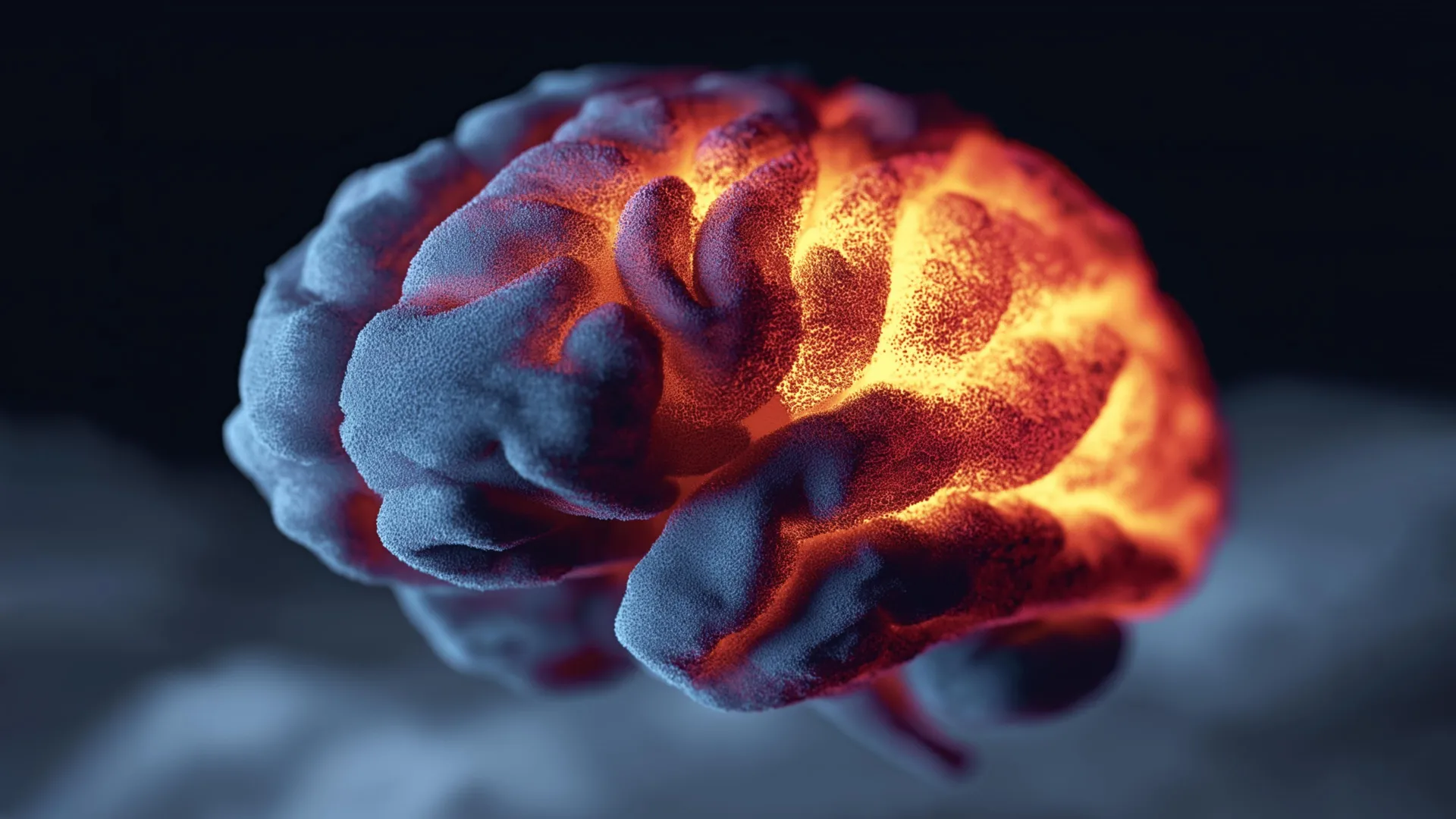
Millions of individuals worldwide grapple with the debilitating reality of chronic nerve pain, a condition that transforms the gentlest touch into an agonizing sensation. For decades, the scientific community has posited that a breakdown in the function of mitochondria – the powerhouses of our cells – within damaged nerves may be a primary instigator of this persistent discomfort. Now, groundbreaking research emerging from Duke University School of Medicine suggests that revitalizing these cellular energy factories could pave the way for an entirely new therapeutic strategy for chronic neuropathic pain.
The findings, meticulously detailed in the prestigious journal Nature, present compelling evidence derived from both human tissue samples and meticulously controlled mouse models. The research team embarked on a mission to determine if replenishing compromised mitochondria could indeed facilitate the recovery of damaged nerve cells. Their investigations yielded significant reductions in pain associated with prevalent conditions such as diabetic neuropathy and chemotherapy-induced nerve damage. Remarkably, in some instances, the pain relief observed persisted for up to an impressive 48 hours, a duration that could substantially improve the quality of life for affected individuals.
This innovative therapeutic avenue diverges from conventional pain management strategies that primarily focus on blocking pain signals. Instead, the Duke researchers propose that their approach targets a fundamental underlying cause of chronic nerve pain: the restoration of the essential energy supply that nerve cells require to maintain proper function.
"By providing damaged nerves with fresh mitochondria, or by stimulating their own production of these vital organelles, we can effectively mitigate inflammation and foster a regenerative environment conducive to healing," explained Dr. Ru-Rong Ji, the study’s senior author. Dr. Ji, who directs the Center for Translational Pain Medicine within the Department of Anesthesiology at Duke School of Medicine, further elaborated on the potential impact of this discovery. "This methodology holds the profound potential to alleviate pain through a mechanism that is fundamentally novel."
The Crucial Role of Healthy Mitochondria in Nerve Recovery
The Duke study’s revelations align with a growing body of scientific understanding regarding the dynamic intercellular transfer of mitochondria. Researchers are increasingly recognizing this phenomenon as an intrinsic cellular support system that may play a significant role in the pathophysiology of a wide array of conditions, extending beyond chronic pain to encompass metabolic disorders like obesity, oncological diseases, and neurological events such as stroke.
At the heart of the Duke researchers’ investigation were satellite glial cells, specialized cells that encircle and provide vital support to sensory neurons. Their study unearthed a previously unrecognized function for these glial cells: the direct donation of healthy mitochondria to sensory neurons. This transfer, the researchers discovered, occurs via minute cellular conduits known as tunneling nanotubes.
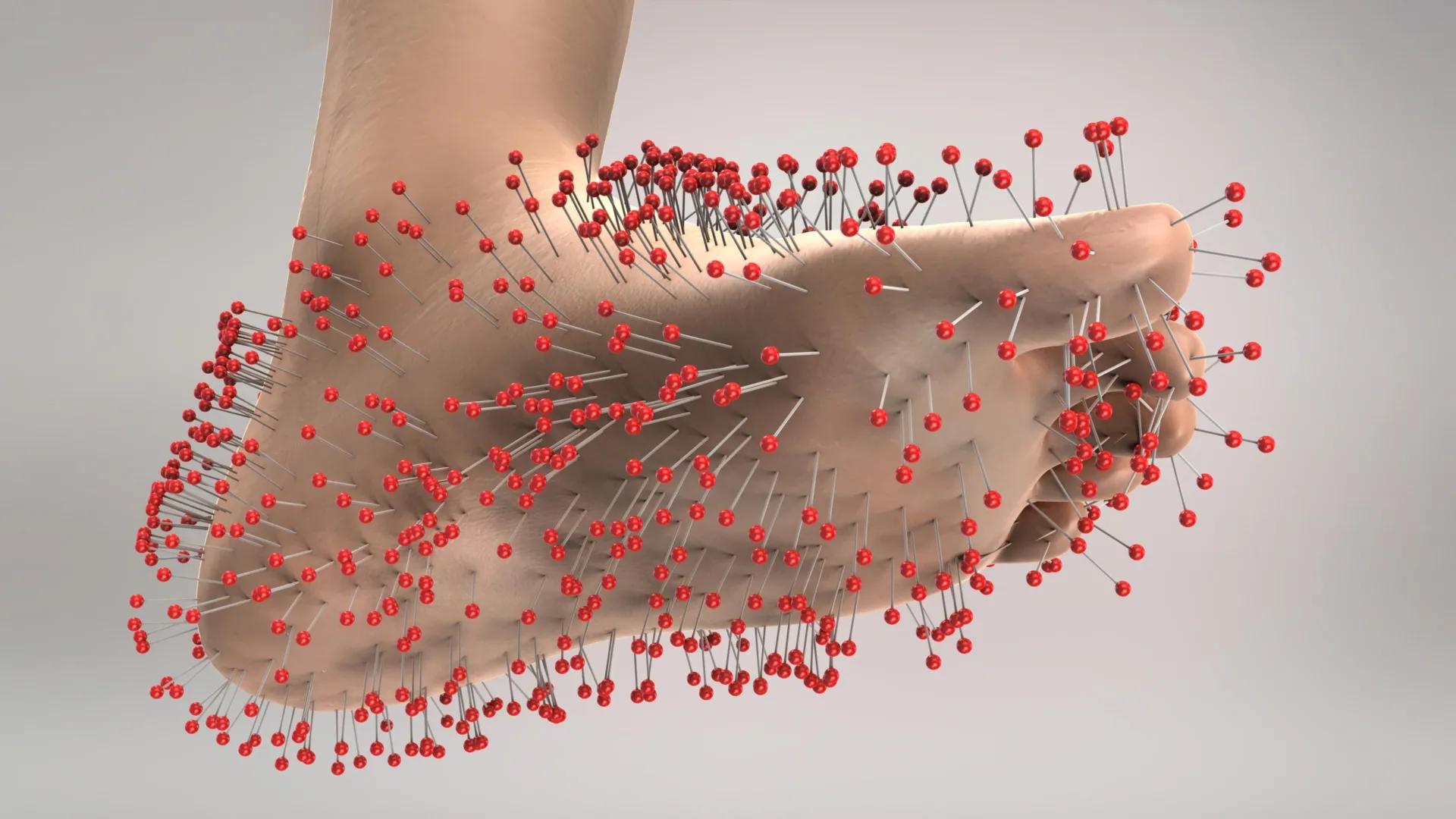
Dr. Ji elucidated the critical importance of this mitochondrial exchange, explaining that when this intricate transfer process falters, nerve fibers begin to degrade. This deterioration, in turn, can manifest as the hallmark symptoms of neuropathic pain, including persistent pain, aberrant sensations like tingling, and a loss of feeling, particularly in the extremities such as the hands and feet, where nerve fibers are most extended.
"Through the sharing of energy reserves, satellite glial cells appear to act as crucial guardians, helping to maintain neuronal integrity and prevent the onset of pain," stated Dr. Ji, who also holds professorial appointments in anesthesiology, neurobiology, and cell biology at Duke School of Medicine. The experimental validation of this hypothesis in their mouse models was striking: an augmentation of this mitochondrial transfer process led to a significant reduction in pain-related behaviors, decreasing by as much as 50%.
Identification of a Key Protein Orchestrating Mitochondrial Transfer
Beyond observing the natural transfer mechanisms, the research team explored a more direct intervention. They administered isolated mitochondria, sourced from both human donors and mice, directly into the dorsal root ganglia. These ganglia are critical clusters of nerve cells responsible for relaying sensory information from the periphery to the brain.
The success of this direct intervention proved highly dependent on the quality of the mitochondria utilized. Healthy donor mitochondria demonstrated a capacity to alleviate pain. Conversely, mitochondria harvested from individuals with diabetes failed to confer any pain-relieving benefits, underscoring the importance of mitochondrial health itself in this therapeutic context.
A significant breakthrough in understanding the mechanics of mitochondrial transfer was the identification of a protein named MYO10. The researchers pinpointed MYO10 as being instrumental in the formation of the tunneling nanotubes, the very structures that facilitate the movement of mitochondria between cells. This discovery provides a crucial molecular target for future therapeutic development.
The collaborative effort behind this landmark study involved lead author Dr. Jing Xu, a research scholar in the Department of Anesthesiology, and long-term collaborator Dr. Caglu Eroglu, a distinguished Duke professor of cell biology renowned for her pioneering work on glial cells.
A Promising New Frontier for Chronic Pain Management
While the current findings represent a substantial leap forward, the researchers emphasize that further investigation is warranted. Future studies will likely involve advanced high-resolution imaging techniques to achieve a more granular understanding of how these nanotubes precisely deliver mitochondria within living nerve tissue.
Nevertheless, the implications of this research are profound. It illuminates a previously underappreciated communication network between neurons and glial cells, a network that could ultimately lead to the development of treatments capable of addressing the root causes of chronic pain, rather than merely suppressing its symptoms. This paradigm shift from palliative care to restorative therapy holds immense promise for millions suffering from intractable neuropathic pain.
Background and Context: The Enduring Challenge of Neuropathic Pain
Neuropathic pain, often described as burning, shooting, or stabbing, arises from damage or dysfunction of the somatosensory nervous system. Unlike nociceptive pain, which signals tissue injury, neuropathic pain is a maladaptive response characterized by aberrant signaling within the nervous system itself. It is estimated that between 7% and 10% of the global population experiences some form of neuropathic pain.
The economic and societal burden of chronic neuropathic pain is substantial, encompassing healthcare costs, lost productivity, and a significant reduction in patients’ overall quality of life. Current treatment options, including analgesics, antidepressants, and anticonvulsants, often provide only partial relief and are associated with considerable side effects. The development of novel therapeutic strategies that target the underlying mechanisms of neuropathic pain has therefore been a long-standing imperative in pain research.
Diabetic neuropathy, a common complication of diabetes mellitus, affects a significant proportion of diabetic patients, leading to nerve damage primarily in the extremities. Chemotherapy-induced peripheral neuropathy (CIPN) is another prevalent and often dose-limiting side effect of various cancer treatments, causing debilitating pain and sensory disturbances. The Duke study’s success in alleviating pain in models of these specific conditions highlights the broad applicability of their mitochondrial restoration approach.
Timeline of Discovery and Future Directions
While the precise timeline leading to this Nature publication is not detailed in the provided text, the research likely represents the culmination of several years of dedicated investigation into glial cell biology, mitochondrial function, and neuropathic pain mechanisms at Duke University. The identification of MYO10 as a key protein, for instance, suggests a systematic approach involving molecular screening and functional assays.
The researchers’ call for further high-resolution imaging indicates that understanding the precise biophysical interactions during mitochondrial transfer is a crucial next step. This could involve employing advanced microscopy techniques like super-resolution microscopy or cryo-electron tomography to visualize the dynamic process of nanotube formation and mitochondrial passage at an unprecedented level of detail.
Furthermore, the translation of these findings into clinical practice will necessitate rigorous preclinical testing in more complex animal models that closely mimic human neuropathic pain conditions. Ultimately, human clinical trials will be required to assess the safety and efficacy of mitochondrial-based therapies for patients.
Potential Impact on Related Fields
The implications of this research extend beyond the immediate realm of neuropathic pain. The understanding of intercellular mitochondrial transfer as a critical support mechanism could have far-reaching consequences for other fields of medicine. For example, impaired mitochondrial function is implicated in the aging process and a host of age-related neurodegenerative diseases. Therapies that enhance mitochondrial health and transfer could potentially play a role in mitigating these conditions.
Moreover, the discovery of MYO10’s role in nanotube formation could open avenues for research into other cellular communication processes mediated by these structures. Tunneling nanotubes have been implicated in the spread of infectious agents and even the transmission of cancer cells, suggesting that understanding and potentially modulating nanotube formation could have therapeutic applications in diverse disease contexts.
Concluding Remarks
The research from Duke University School of Medicine represents a significant advancement in our understanding of chronic nerve pain and offers a tangible pathway toward innovative treatment strategies. By focusing on the fundamental energetic health of nerve cells and the intricate mechanisms of intercellular support, scientists are moving closer to providing genuine relief for individuals suffering from debilitating neuropathic pain. The journey from laboratory discovery to patient bedside is often long, but the promise held within the concept of restoring cellular powerhouses offers a beacon of hope for a future with less pain.