Alzheimer’s disease, a relentless neurodegenerative condition, casts a long shadow over millions of lives, with an estimated 7.2 million Americans aged 65 and older affected, according to the Alzheimer’s Association. The current diagnostic landscape primarily relies on measuring the levels of two key proteins – amyloid beta (Aβ) and phosphorylated tau (p-tau) – in blood or cerebrospinal fluid. While these established biomarkers have been instrumental in advancing our understanding and diagnosis of Alzheimer’s, a growing body of research suggests they may not capture the full spectrum of the disease’s earliest biological transformations. This limitation has spurred a quest for more sensitive and comprehensive diagnostic tools, a quest that may have taken a significant leap forward with the groundbreaking work from researchers at Scripps Research.
In a pivotal study published on February 27, 2026, in the esteemed journal Nature Aging, scientists unveiled a novel blood test that shifts the diagnostic paradigm from quantifying protein amounts to analyzing protein structure. This innovative approach focuses on the intricate ways proteins fold within the bloodstream, revealing that specific structural alterations in just three plasma proteins are profoundly linked to an individual’s Alzheimer’s status. The implications of this discovery are far-reaching, offering the potential for earlier and more accurate identification of individuals with Alzheimer’s disease and even its precursor, mild cognitive impairment (MCI).
The Protein Folding Conundrum: A New Frontier in Alzheimer’s Research
For decades, the pathological hallmarks of Alzheimer’s disease have been widely understood as the aberrant accumulation of amyloid plaques and tau tangles within the brain. These protein aggregates are considered primary drivers of neuronal damage and cognitive decline. However, the scientific community is increasingly embracing a more holistic view of neurodegenerative diseases, recognizing that they may stem from a systemic breakdown in proteostasis – the intricate cellular machinery responsible for maintaining protein health. Proteostasis encompasses the processes of protein synthesis, proper folding, refolding of damaged proteins, and the clearance of misfolded or aggregated proteins.
As the human body ages, the efficiency of this vital proteostasis system naturally diminishes. This age-related decline renders proteins more susceptible to misfolding during their production or maintenance phases. The hypothesis underpinning the Scripps Research study was elegantly simple yet profoundly significant: if the proteostasis system is compromised in the brain, leading to protein misfolding associated with Alzheimer’s, then similar structural deviations might also manifest in proteins circulating throughout the body’s bloodstream. This premise laid the groundwork for an investigation into the "structural signature" of blood proteins as a potential diagnostic window into neurological health.
Unraveling Structural Signatures: A Deep Dive into Blood Proteins
To rigorously test their hypothesis, the research team meticulously analyzed plasma samples collected from a cohort of 520 participants. This cohort was strategically divided into three distinct groups: cognitively normal individuals, those diagnosed with mild cognitive impairment (MCI), and patients with a confirmed diagnosis of Alzheimer’s disease. The selection of participants across this spectrum of cognitive function was crucial for establishing clear correlations between protein structural changes and disease progression.
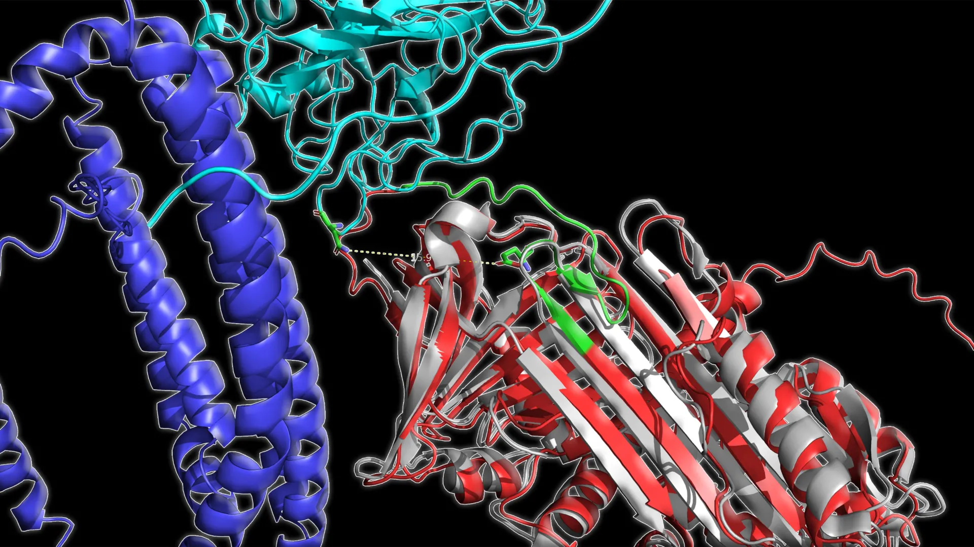
The researchers employed advanced mass spectrometry techniques, a powerful analytical tool capable of dissecting the molecular composition and structure of proteins. This method allowed them to precisely map the accessibility of specific regions within proteins, essentially determining which parts were exposed to the surrounding environment and which were buried. Changes in this exposure pattern serve as a direct indicator of alterations in protein structure. Following the mass spectrometry analysis, the team harnessed the power of machine learning algorithms. These sophisticated computational tools were instrumental in identifying subtle yet consistent patterns within the structural data that were uniquely associated with each disease stage.
The results of this comprehensive analysis were striking. A clear and discernible pattern emerged across all participant groups. As Alzheimer’s disease advanced, certain plasma proteins exhibited a progressive decrease in their structural "openness" – a sign of altered folding. This discovery was particularly significant because these structural changes proved to be far more informative in distinguishing disease stages than simply measuring the concentration of these proteins, a limitation of current diagnostic approaches.
The Trio of Proteins: Key Players in Alzheimer’s Progression
Among the vast array of proteins present in plasma, a select group of three demonstrated an exceptionally strong correlation with Alzheimer’s disease status. These proteins, each with distinct physiological roles, collectively painted a compelling picture of the disease’s impact on protein integrity.
The first protein identified was C1QA, a crucial component of the immune system, specifically involved in the complement cascade, which plays a role in immune signaling and the clearance of cellular debris. Dysregulation of immune responses has been increasingly implicated in the pathogenesis of Alzheimer’s disease, making C1QA a significant marker.
The second protein, clusterin, is a multifaceted chaperone protein known to be involved in a variety of cellular processes, including protein folding, stabilization of other proteins, and the clearance of misfolded proteins and amyloid aggregates. Its involvement in amyloid removal makes it a particularly relevant player in the context of Alzheimer’s pathology.
The third protein, apolipoprotein B, is a major component of low-density lipoproteins (LDLs) and plays a critical role in transporting fats (cholesterol and triglycerides) in the bloodstream. It also contributes to the health and integrity of blood vessels. Emerging research has highlighted the complex interplay between vascular health and neurodegenerative diseases, suggesting that alterations in lipid metabolism and transport could have downstream effects on brain function and disease progression.
Casimir Bamberger, a senior scientist at Scripps Research and co-author of the study, expressed his astonishment at the findings, stating, "The correlation was amazing. It was very surprising to find three lysine sites on three different proteins that correlate so highly with disease state." Lysine sites are specific amino acid residues within proteins that can be modified, and changes in their accessibility can reflect structural alterations.
The power of this three-protein structural signature was further quantified. Researchers were able to classify participants into cognitively normal, MCI, or Alzheimer’s disease categories with an impressive overall accuracy of approximately 83%. When comparing just two groups directly, such as cognitively normal individuals against those with MCI, the accuracy surged to over 93%. This level of diagnostic precision is a significant advancement over current methods, which often struggle with early and definitive differentiation.
Tracking the Disease: Temporal Stability and Predictive Power
A critical aspect of any new diagnostic test is its reliability and consistency over time. The researchers addressed this by rigorously testing their three-protein model in independent cohorts of participants. Furthermore, they analyzed blood samples that had been collected months apart from the same individuals. The results were highly encouraging. The model maintained its predictive power, identifying disease status with approximately 86% accuracy in repeat tests conducted months apart.
Crucially, the structural score derived from these three proteins not only reflected the current disease status but also appeared to track changes over time. This suggests that the test could potentially monitor the progression of Alzheimer’s disease, a capability that is currently limited with existing biomarkers. Moreover, the structural score exhibited a strong relationship with results from standard cognitive tests, further validating its clinical relevance. The study also found a more moderate, yet still significant, association between the structural score and MRI measurements of brain shrinkage, a common indicator of neurodegeneration in Alzheimer’s.
These comprehensive findings strongly suggest that analyzing the structural characteristics of blood proteins could serve as a valuable complement to existing amyloid and tau-based diagnostic tests. By focusing on structural changes intrinsically linked to the underlying biological mechanisms of Alzheimer’s, this novel method holds the potential to enhance the identification of disease stages, provide a means to monitor disease progression, and critically, to evaluate the efficacy of therapeutic interventions.
The Promise of Early Intervention: A New Era for Therapeutics
The implications of a highly accurate and accessible early diagnostic tool for Alzheimer’s disease are profound, particularly in the realm of therapeutic development. "Detecting markers of Alzheimer’s early is absolutely critical to developing effective therapeutics," emphasized senior author John Yates, a professor at Scripps Research. "If treatment can start before significant damage has been done, it may be possible to better preserve long-term memory."
The current therapeutic landscape for Alzheimer’s disease is largely focused on managing symptoms or, in some cases, targeting the underlying protein pathologies. However, the effectiveness of many treatments is significantly hampered by the fact that diagnoses are often made when substantial neuronal damage has already occurred. By enabling interventions at the earliest detectable stages of the disease, when pathological changes are still nascent and the brain retains greater resilience, there is a much greater hope of slowing or even halting disease progression. This could translate into preserving cognitive function for longer periods, significantly improving the quality of life for patients and their families.
Pathways to Clinical Adoption and Future Horizons
While the results of this study represent a monumental step forward, the path to widespread clinical adoption requires further validation. The researchers acknowledge that larger, longitudinal studies with extended follow-up periods are essential to confirm these findings in diverse populations and to solidify the test’s reliability in real-world clinical settings.
Beyond Alzheimer’s disease, the Scripps Research team is actively exploring the broader applicability of their structural profiling method. The underlying principle of analyzing protein misfolding and structural aberrations is not exclusive to Alzheimer’s. This innovative technique may hold promise for the early detection and monitoring of other complex diseases, including Parkinson’s disease, other forms of dementia, and even certain types of cancer, many of which are also characterized by disruptions in proteostasis.
The study, titled "Structural signature of plasma proteins classifies the status of Alzheimer’s disease," lists Ahrum Son, Hyunsoo Kim, and Jolene K. Diedrich from Scripps Research as authors, alongside Heather M. Wilkins, Jeffrey M. Burns, Jill K. Morris, and Russell H. Swerdlow from the University of Kansas Medical Center, and Robert A. Rissman from the University of California San Diego. This collaborative effort underscores the multi-institutional commitment to tackling the challenges of neurodegenerative diseases.
Financial support for this pivotal research was provided by the National Institutes of Health, through grants RF1AG061846-01, 5R01AG075862, P30AG072973, and P30-AG066530, highlighting the critical role of federal funding in advancing cutting-edge scientific discovery. The successful development and implementation of this novel blood test could usher in a new era of Alzheimer’s diagnosis and treatment, offering a beacon of hope for millions affected by this devastating disease.